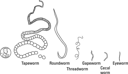
A parasite is a living organism that depends upon a living host for survival. It lives either inside or outside the host and thereby causes discomfort or inefficiency in the productive life of the host. Those that live inside the host are called internal parasites and those outside are referred to as external parasites.
The Different Internal Parasites of Ruminants
Most of the internal parasites are either worms or flukes which are collectively referred to as helminths. They live in the lungs, liver, stomach, and intestines of animals.
1. Roundworms
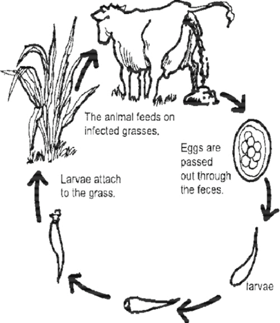
Roundworms found in the gastrointestinal tract are called nematodes. The organism that causes the most offensive damage is Haemonchus contortus or twisted worm. Others include Ostertagia (brown stomach worm), Trichostrongylus asei, and Trichostrongylus vitrinus. They vary in their size from tiny thread-like structures of about 5 mm long to over 300 mm.
The life cycle of the roundworm is shown in the image above. The worms mate and lay eggs inside the abomasum of the ruminants which are expelled along with the dung. They hatch and develop to the larva stage on the pasture where they are picked up by grazing animals.
The larva cannot survive harsh and dry weather but thrive well in moist and warm weather even for up to two years. They develop into mature roundworms within twenty-one days from the time the egg is laid.
In the intestine, the worms damage the inner lining so that blood, nutrients, and water are lost in the feces or urine. Infestation of worms is often accompanied by diarrhea, dehydration, and loss of appetite. The implication of this is that nutrients in the feed will be poorly utilized for growth and production purposes.
While sheep and goats suffer from the same species of roundworm, cattle has a different species. Hence sheep and goats can graze together without fear of cross infestation.
Worms are often controlled by developing programs that will match the seasonal occurrence. Routine drenching with chemicals such as benzimidazole, levamisole, and organophosphate is necessary.
The herd should be de-wormed about 6 to 8 weeks after they might have started grazing during the rainy season and repeated two weeks after. This action is repeated shortly before late rains and the onset of the dry season. The herd must not be allowed to graze infested pasture.
Read Also Effect of Tropical Climate on Animal Parasites, Vectors and Diseases
2. Lungworms
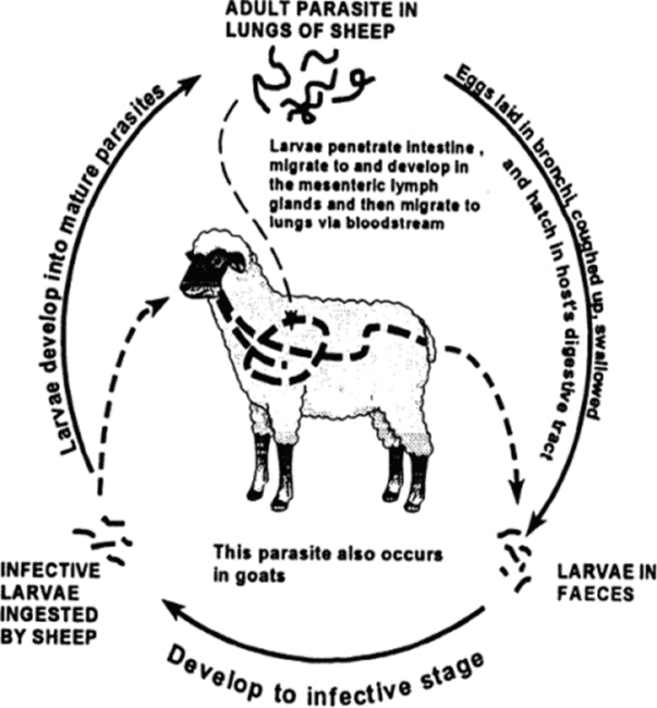
Lungworm is caused by Dictyocaulus filaria or Dictyocaulus viviparus. They infect the lungs of the animal causing considerable damage to them and the bronchial tube which leads to coughing.
The symptoms are irregular breathing, coughing associated with worms, and blood-stained discharge being expelled from the mouth. The infected animal tends to stand in distress on pasture and take little or no interest in grazing. There is also loss of body condition.
The life cycle of lungworms is shown in the image above. The adult lays eggs in the air passages of the lungs where they live. The eggs are coughed up into the back of the throat and swallowed. The eggs then pass through the alimentary canal of the animal and hatch into immature larvae which are expelled along with the dung onto the pasture.
Animals that are grazing easily pick up the larva, and pass them down through the alimentary canal where they infest the walls, and find their way into the blood vessels.
They are subsequently carried to the heart and the lungs. On getting to the lungs, they bore through the tissues of the tiny air space causing a lot of damage. They are at this stage capable of laying eggs which are coughed out to the back of the throat and the cycle is re-started.
Lungworms are controlled by the use of appropriate anthelmintics as may be advised by the veterinarian. However, the use of live oral vaccine as a routine immunization before turning the animals to pasture has been reported in Northern Europe only. This is very important for young animals like calves.
3. Tapeworms
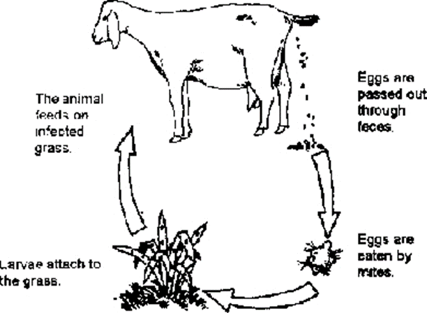
Tapeworms (gestoda) belong to the phylum Platyhelminthes and are related to the flukes (trematodes). They have segmented bodies that are tape-like and also host-specific. They infect both animals and humans. The adult tapeworm causes fewer problems, especially in animals under poor plane of nutrition. They are also non-pathogenic. However, the larva of some species travel to the brain or the spinal cord and cause a nervous disorder called coenurosis.
The life cycle of tapeworm is shown in the image above. When a dog eats the carcass of infected sheep or ruminants, the cysts develop into tapeworms inside the intestine of the dog. The matured worm lays eggs that pass along with the dog’s feces.
Grazing ruminants pick the eggs while on pasture which hatches and move into the blood vessels and eventually find their way to the brain or the spinal cord. If a dog eats the carcass of infested ruminants, the cycle is restarted. For a control measure and to break the cycle, dogs must not be allowed to eat the carcasses of infected animals. Benzimidazole can be used for treatment.
4. Trypanosomiasis
This is a protozoan infection that affects both humans and animals, especially ruminants. It is caused by trypanosome parasites which could be trypanosoma vivax, Trypanosoma brucei, or Trypanosoma evansi. They invade the bloodstream and cause symptoms such as recurrent fever (with about twelve days in between), anemia, discharge from the eyes, nervous signs, and loss of condition. The animal may be infected for months before it dies finally.
The vector for this disease is tsetse fly which is very prevalent in sub-Saharan Africa. There exist some wild animals that are infected but show no symptoms and are therefore carriers of the parasites. These wild animals are buffaloes, giraffes, and warthogs. Tsetse flies sucks blood from these wild animals and thereby maintains the cycle of the parasites. Human beings are also susceptible when they are bitten by these vectors.
Trypanosomiases are treated by the use of trypanocidal drugs such as diminazene aceturate, and homidium chloride. Because of the prevalence, prophylactic drugs are available and used by injection to last for about three months. Such drugs include isometamidium chloride and quinapyramine pro salt. Regular spraying of the environment with insecticides to kill tsetse flies is another control measure apart from prophylaxis.
5. Liver Fluke
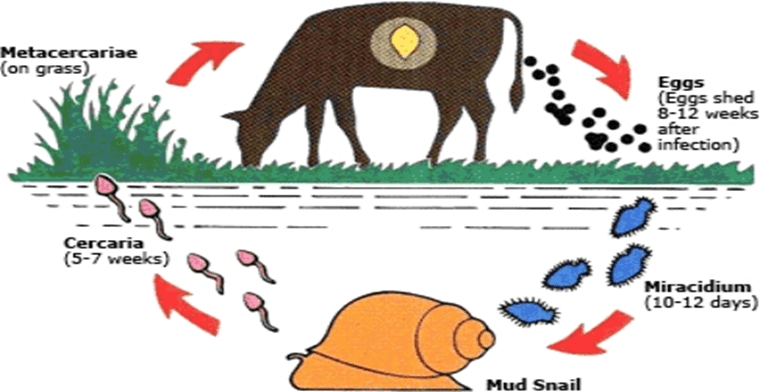
This is one of the most widely distributed and harmful parasites that affect cattle sheep and goats. They are caused by Fasciola hepatica or Fasciola gigantica of the trematodes. The disease symptoms include paleness of the eyelid and the gum, pot-bellied condition, appearance of soft watery swelling under the jaw, weakness, anemia, and loss of condition. When the carcass of infested ruminants is posted, the flukes are found in the liver if opened. It causes a disease called schistosomiasis.
While in the sheep, the adult fluke lays eggs in the bile duct. The eggs are passed into the intestine and expelled along with the dung onto the pasture. Here they can stay up for six months if it is in a wet environment or they die if on dry land. They hatch into miracidium (after about nine days to eight weeks) which swims and flows with streams or brooks or any water in the drains around.
The miracidium is picked up by the water or mud snails (Limmaea truncatula) and after about seven weeks they develop and produce another form called cercariae. An average of 1000 cercariae is produced from one miracidium. The cercariae move and attach themselves to the leaves of the plants or grasses around where grazing ruminants pick them up in the encysted form.
They migrate into the liver of the animal via the blood vessels and develop into liver fluke after about six weeks. The flukes begin to lay eggs after another six weeks in the ruminant host which are again expelled along with the dung and the cycle continues as shown in the image above.
Liver fluke is treated with benzimidazole and salicylanilides. It is controlled by the elimination of the intermediate host- mud snail. This is achieved by spraying the streams, brooks, and every drain around the grazing pasture regularly during the wet season.
6. Coccidia
This is a protozoa disease caused by the Eimeria spp of bacteria. The parasites live in the intestine of the animal. The main symptom is blood-stained diarrhea where ruminants especially young ones are raised intensively.
It can be treated by the administration of sulpha drugs and antibiotics. Control measures are by preventing the animals from eating feeds contaminated with feces.
In summary, parasites are of economic importance in the rearing of ruminants for production purposes. Parasites can be internal or external but live on their host to survive by damaging the physiological conditions of the host. The internal parasites include worms, trypanosomiasis, liver flukes, and coccidia.
Read Also How to Convert Organic Waste (Composting) into Compost for Gardening and Agriculture

