

Microbiology, the study of microscopic (very small) forms of life, has transformed our understanding of the world. Throughout most of history, people had no knowledge of the minute organisms that exist everywhere, and the implications of this ignorance were huge.
Infectious disease could not be understood, prevented, or cured prior to the discovery of microorganisms. Modern infectious disease medicine is built upon a solid understanding of microbiology. In this article, we shall examine the meaning of microbiology, the role of microorganisms, and characteristics of microbes.
Defining Microbiology
Microbiology is the study of tiny or minute living organisms that are not directly visible to the naked eye but are only visible with the use of microscopes. Microbiology can also be defined as a specialized area of biology that deals with living things too small to be seen without magnification. These microorganisms are collectively called microorganisms or microbes. They can be divided into five groups:
| Plural | Singular |
|---|---|
| Bacteria | Bacterium |
| Fungi | Fungus |
| Viruses | Virus |
| Algae | Alga |
| Protozoa | Protozoan |
Microbiology studies the diversity, evolution, and beneficial and harmful roles of microorganisms to mankind and their environment as a whole. This is because microorganisms affect all forms of life on earth.
Roles and Uses of Microorganisms in Agriculture and Beyond
Microbiology helps humans understand the basic life processes on earth and enables them to utilize this knowledge to their benefit. The following are some of the roles of microorganisms, which may be beneficial and harmless or harmful and pathogenic to humans, plants, animals, and their environment.
Microorganisms in Agriculture
Microorganisms can cause diseases in plants and animals, thereby bringing about economic losses to farmers and the nation as a whole. Ruminant animals also benefit from the activities of microbes living in their digestive systems.
These organisms help the animals digest cellulose-rich diets. The association between leguminous plant root nodules serves to convert atmospheric nitrogen to ammonia, which the plant can readily use for growth, thus reducing the need for expensive and polluting nitrogen fertilizers.
Microorganisms in Food Production
Food is an essential medium or material rich in nutrients that can support life, including that of microbes. This makes it easy or possible for organisms to cause food poisoning when they contaminate such foods. When this happens, the food decays, gives off flavors, and becomes unsuitable for consumption.
Food spoilage causes economic losses to humans. Fermentation is another important activity in which microbes are useful. They are used in the fermentation of alcoholic beverages (e.g., beer), cheese, yogurt, and in baking, where yeast is used to raise dough.
Microbes can also use food as a material for transmitting foodborne diseases. The food material is referred to as a ‘vehicle of infection.’ Water can serve as a vehicle for the transfer of cholera; salad can serve as a vehicle of transfer for food poisoning.
Microorganisms in Nutrient Recycling
Microbes are useful in the recycling of some nutrients in nature, e.g., the carbon, nitrogen, and oxygen cycles that take place in nature. The activities of some microbes also produce natural gases such as methane. Some other microbes are used on agricultural waste, domestic waste, and some industrial waste to generate biofuels such as ethanol and methane.
For example, ethanol is produced by microbial fermentation of glucose from sugarcane or corn starch and is the major motor fuel in some countries, such as Brazil.
Microorganisms are also natural cleaning agents in their environment, with bioremediation abilities. For example, some microbes have the ability to clean up spilled oil, pesticides, or toxic environmental pollutants by degrading such materials.
Microorganisms in Medicine
A large number of microbes cause diseases in animals, plants, and humans. These organisms may use vectors (i.e., living organisms, e.g., flies, mosquitoes, and cockroaches) or vehicles (i.e., inanimate objects) to transmit diseases from one person to another.
The knowledge of the harmful roles of microbes has helped in the identification, development, treatment, and prevention of such diseases, for example, through the use of antibiotics and improved sanitary and public health practices. The microbes that cause diseases are called pathogens.
Read Also: Goats Farming Complete Practical Guide
Microorganisms in Biotechnology
Some microbes are used in the manufacturing of products of commercial value, e.g., vaccines, antibiotics, and hormones. They are also useful in the food industry in the production of fermented products on a large scale. Biotechnology is the use of genetically modified microbes to synthesize products, e.g., human insulin is produced by modified bacteria.
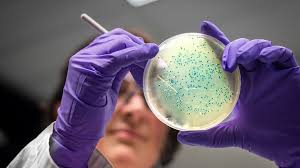
Microscopic Examination of Microorganisms
This is usually the first step in the identification of an organism. Due to the very small size of these organisms, there is a need to magnify them in order to view and study their internal organelles.
1. Types of Microscopes
Microscopes are instruments used to magnify objects. There are two types:
i. Light Microscope: Its name is derived from the fact that it uses a light source for illumination. The light may be natural sunlight or electricity. The different types of light microscopes include:
- Bright field
- Dark field
- Phase contrast
- Fluorescence
- Confocal scanning laser microscope
ii. Electron Microscope: This uses electron beams and is usually used to view viruses and large molecules. The major difference between it and the light microscope is their source of illumination. Examples of electron microscopes are:
- Transmission electron microscope
- Scanning electron microscope
2. Staining Techniques for Microorganisms
This involves the application of different stains and dyes on prepared specimens to enhance their visibility under the microscope. Staining is mainly done because the cytoplasm of most organisms is colorless, making it difficult to observe the organism.
Therefore, staining is done to enhance visibility, emphasize specific morphological features (structural features), and preserve stained specimens for future use. Different staining techniques include:
- Gram staining technique
- Spore staining technique
- Capsule staining
- Flagella staining
- Acid-fast staining
Before staining, the specimen must be subjected to some treatments, such as:
- i. Smear Preparation: This involves spreading a thin film of the cell suspension on a glass slide and allowing it to dry.
- ii. Fixation: This involves passing the air-dried smear through a flame and allowing it to gently heat up. The heating kills and fixes the cell on the slide; it also inactivates enzymes that may change the morphology of the cell. It also toughens the cell so that it does not change during staining and observation. Apart from the use of heat, chemicals can be used in fixation, e.g., ethanol, acetic acid, etc.
- iii. Staining: This is done by the application of dyes to the specimen to impart colors through a chemical reaction that allows the dyes to bind to the cell.
There are two major groups of stains used: basic dyes and acid dyes.
| Basic Dyes | Acid Dyes |
|---|---|
| Crystal violet | Eosin dye |
| Methylene blue | Congo red |
| Basic fuchsin | Indian ink |
| Safranin | Black stain |
| Malachite green | Rose Bengal |
The basic dyes are positively charged and are attracted to the negative charge of the cytoplasm, while the acid dyes are negatively charged and are repelled by the negative charge of the cytoplasm, thus the stain gathers around the edge of the cell. Staining methods can also be simple or differential.
3. Gram Staining Technique
It is an example of differential staining. It was developed in 1884 by Christian Gram. It is an important staining technique in microbiology used to differentiate bacteria into gram-positive and gram-negative based on the cell wall of the organism.
4. Procedure for Gram-Positive and Gram-Negative Bacteria
- Make a smear of the specimen on a clean glass slide and allow it to dry.
- Heat fix the smear by passing through a flame 2-3 times.
- Flood the slide with crystal violet and allow it to dry.
- Wash off excess dye with water.
- Add Gram’s iodine and wait for 60 seconds.
- Flood the slide with alcohol or acetone and leave it for 10 seconds.
- Wash off excess with water.
- Counterstain with carbon fuchsin or safranin.
- Wash off excess with water.
- Blot dry the slide and observe under the microscope.
5. Observation and Result of Gram Staining
If a purple or violet color is seen, the bacterium is a gram-positive bacterium. This is due to the ability of the cell to retain the primary dye and resist decolorization. If a pink or red coloration is observed, the organism is a gram-negative bacterium. This is because alcohol is able to decolorize the primary stain, making it possible for the cell to obtain and retain the counterstain.
6. Roles of the Different Stains or Reagents Used for Gram Staining
i. Crystal Violet: This is the primary stain, and it colorizes the specimen purple or violet.
ii. Gram’s Iodine: This is the mordant; it enhances the interaction between the cell and the dye so that the cell can take up the color more strongly.
iii. Alcohol or Acetone: This is the decolorizer, and it determines if the cell is going to be decolorized or not of the primary stain.
iv. Safranin or Carbon Fuchsin: This is the counterstain that gives the pink or reddish color to the cell after it has been decolorized.
| Crystal Violet | Gram’s Iodine | Alcohol or Acetone | Safranin or Carbon Fuchsin | |
|---|---|---|---|---|
| Gram + Bacterium | Purple | Purple | Purple | Purple/Violet |
| Gram – Bacterium | Purple | Purple | Cell is decolorized | Red/Pink |
Characteristics of Microbes
Microbes are not all alike; they vary in size, shape, structure, and function, but they share a common feature: they are microscopic. They are usually unicellular in nature, and most are classified under the kingdom Protista. All living things may be divided into two groups:
1. The Prokaryotes
They are simple organisms composed of single cells having no membrane-bound intracellular organelles. They lack a well-developed nucleus, and their ribosomes are free in the cytoplasm. Examples include bacteria and blue-green algae.
2. The Eukaryotes
They possess a nucleus with a well-defined nuclear membrane. They also have membrane-bound organelles, such as mitochondria, in the cytoplasm. Examples are protozoa and fungi.
| Prokaryotes | Eukaryotes | |
|---|---|---|
| Nucleus | Not well defined | True nucleus |
| Nuclear Membrane | Absent | Present |
| Chlorophyll | When present, it is dissolved in cytoplasm | When present, it is found in the chloroplast |
| Cell Wall | Usually made of peptidoglycan | Contains cellulose or chitin |
| Reproduction | Asexual method | Both sexual and asexual |
| Organelle | Few external organelles | Many internal organelles |
| Chromosome | One | Many |
| Ribosome | Small | Large |
| Mitosis | No evidence | Takes place |
Read Also: Introduction to Ruminant Animals Production
Morphological Characteristics of Microorganisms
1. Protozoa: They are unicellular animals that live in aqueous environments, such as ponds; the majority are free-living and harmless, e.g., amoeba. While a few are parasitic and cause diseases, for example, Entamoeba histolytica and Plasmodium sp.
2. Algae: All algae manufacture their food through photosynthesis. They may be microscopic or macroscopic in size, e.g., seaweeds. They are usually free-living and harmless, e.g., in the green slime on the surface of ponds and algae.
Algae are pigmented organisms; they are eukaryotic but lack a well-developed vascular system. They have chlorophyll and reproduce asexually and sexually. They also store their carbohydrates as glucose, e.g., Euglena.
3. Viruses: They are the smallest of all microbes, e.g., bacteriophage. They are obligate intracellular parasites that depend on their host for all their metabolic activities; thus, outside their living host, they are metabolically inactive and are often referred to as crystallizable chemicals.
When inside the host, they redirect the activities of the host cells to replicate themselves. Viruses do not grow on artificial media. They contain either DNA or RNA but never both.
The DNA or RNA is enclosed in a layer of protein called a capsid. Viruses are acellular organisms, i.e., they lack cellular organization. They cause diseases in both plants and animals. Viruses also infect bacteria, and such bacteria are called bacteriophages.
4. Fungi: They are eukaryotic organisms that lack chlorophyll and flagella. They may exist as multicellular filamentous hyphae or unicellular. The unicellular fungi are referred to as yeast. They are unicellular because they exist as single-cell organisms, although a large mass of yeast can be seen by the naked eye; they are individually microscopic.
They are found on leaves, flowers, and secretions of leaves; they can also be found on the skin and alimentary canal of humans. Some yeast is pathogenic to humans, animals, or plants.
They are also useful in several industries to produce wine, beer, and bread because they are able to ferment sugars to produce CO2 and alcohol. Examples of yeast include Saccharomyces cerevisiae and Candida albicans.
5. Molds: The multicellular filamentous cells are called molds. They can be found on damp newspapers, old leather, decaying wood, rotten food, or walls, etc. They are called multicellular because they are composed of many cells joined together. They are able to break down organic materials into simple forms.
They are often involved in food spoilage, e.g., Rhizopus sp. causes spoilage in bread. They also cause diseases, e.g., ringworm. Molds are also useful when they grow on some foods, e.g., the ripening of cheese.
Molds are made up of many threads called hyphae. A collection of these hyphae is called mycelium. Reproduction may be asexual or sexual. Examples include Penicillium sp. and Aspergillus sp.
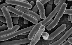
6. Bacteria: They are prokaryotic organisms that reproduce by binary fission. They are unicellular organisms with various nutritional requirements, and some may live in an oxygen-rich environment or an oxygen-deficient environment. Bacteria that live in an oxygen-rich environment are called aerobic bacteria, and those that live in an oxygen-deficient environment are called anaerobic bacteria.
Growth may also occur at different temperatures depending on the type of bacteria and where it is found. They may be found in the soil, plants, water, or humans. The shape and arrangement of bacteria also differ.
Bacteria may be gram-positive or gram-negative, depending on their reaction to gram staining. They may also possess additional structures such as flagella, capsules, and endospores. The cell wall of bacteria is made of peptidoglycan, and it gives shape to the bacteria. It can be pathogenic to humans, plants, and animals.
7. Mycoplasma: These are bacteria that lack a cell wall because they cannot produce peptidoglycan. They are pleomorphic, i.e., they have no rigid shape; their sizes vary, and their genetic material is the smallest among prokaryotes. An example is Mycoplasma sp.
8. Rickettsiae: They are intracellular parasites believed to be between bacteria and viruses due to their small size and intracellular nature. They may be rod-shaped or cocci, but some may be pleomorphic (no shape), e.g., Rickettsia typhi, Rickettsia prowazekii.
9. Cyanobacteria (or Blue-Green Algae): They appear green due to the pigment they contain. They are photosynthetic bacteria. They may exist as unicellular or filamentous forms. Their cell wall is similar to that of gram-negative bacteria.
They are believed to be between bacteria and algae because their photosynthetic system closely resembles that of algae. Examples of cyanobacteria include Nostoc, Anabaena sp., and Oscillatoria.
Structure of Bacteria
All bacteria have the same internal structures regardless of their individual shape. This may include the following:
- Rigid Cell Wall
- Cell Membrane
- Double-Stranded DNA
- Ribosomes
- Cytoplasm
- Mesosome
- Storage Granules and additional structures such as:
- Flagellum
- Capsule
- Endospores
The outer layer of the bacterial cell is called a cell wall. It is rigid, strong, and maintains the shape of the organism. It also acts as a barrier to certain compounds. The cell wall is made of a peptidoglycan layer, which is thicker in gram-positive than in gram-negative bacteria.
The cell membrane controls the entry and exit of all substances and surrounds structures within the cell cytoplasm. The DNA is the nuclear material that carries the genes of the cell, which determines the nature of the bacterium.
The cytoplasm is made of water, sugar, amino acids, salt, etc., and supports the ribosomes, glycogen, and lipids. The mesosome is found in many gram-positive bacteria and is important in cell division.
Additional Bacterial Structures
1. Flagella: A flagellum is a thread-like appendage attached to the cell membrane. It is a locomotion appendage responsible for the mobility of the bacterium. The pattern of distribution of flagella in bacteria differs. There are four different distribution patterns:
i. Monotrichous Distribution: This is observed in bacteria that have a single flagellum located at one end of the bacteria, e.g., Vibrio cholerae.
ii. Amphitrichous Distribution: This is when one flagellum is seen attached to each end of the bacteria, e.g., Pseudomonas aeruginosa.
iii. Lophotrichous Distribution: These are bacteria that have a cluster or tuft of flagella at one end or at both ends, e.g., Alcaligenes faecalis.
iv. Peritrichous Distribution: This occurs when flagella are evenly spread over the whole outer surface of the bacterium, e.g., Proteus mirabilis.
2. Capsule: This is a layer of gelatinous material produced by the bacterial cell itself, which adheres to the outside of the bacterial cell. The function is to protect the bacterium against destruction. Bacteria that have a capsule are said to be encapsulated. Examples are Bacillus subtilis and Bacillus anthracis.
3. Endospores: These structures are commonly referred to as spores. They are spherical or oval structures formed within a vegetative bacterial cell. They contain enough materials to sustain them when they are released from the parent cell.
A mature spore can remain in a dormant state for a very long period because they have extraordinary resistance to heat, cold, and chemicals. They can survive in dust, vegetation, or soil for several months or years until they find a suitable environment for reproduction. The position of spores in cells includes the following:
i. Central Spore: This occurs when the spore is located at the center of the bacterium, e.g., Bacillus anthracis.
ii. Subterminal Spore: This is seen when the spore is located toward one end of the bacteria. Sometimes, the diameter will be greater than that of the cell, thereby causing the cell wall to bulge, forming a spindle shape, e.g., Clostridium botulinum.
iii. Terminal Spore: This is when the spore is located at one end of the bacterial cell. If the diameter of the spore is greater than that of the bacteria, it bulges and gives a drumstick appearance, e.g., Bacillus subtilis, Clostridium tetani.
Classification of Bacteria Based on Microscopic Morphology
1. Bacterial Shape: When bacterial specimens are stained and viewed under the microscope, different shapes and arrangements can be observed. This can be used for preliminary identification.
i. Coccus: These are spherical or near-spherical cells. There are varieties of arrangements for cocci cells.
ii. Cluster Arrangement: When there is a cluster arrangement, they are said to be staphylococci, e.g., Staphylococcus aureus.
iii. Chain Arrangement: This is when the cocci cells are arranged in chains. They are said to be streptococci, e.g., Streptococcus pyogenes.
iv. Pair Arrangement: When this occurs, they are said to be diplococci, e.g., Streptococcus pneumoniae.
v. Bacillus: This is a rod-shaped cell. It may have rounded ends or large square ends.
vi. Vibrio: These are comma-shaped rod cells.
vii. Spirillum: These are spirally shaped curved rods.
Apart from shape, other microscopic morphological classifications that are used include:
- Colony shape
- Colony margin or edge (smooth, serrated)
- Colony elevation (flat, raised on the agar)
- Colony surface (wrinkled, smooth, glittering, dull)
- Colony pigmentation (color)
- Odor (sweet, decay, fruity)
Growth Pattern of Bacteria in Liquid Broth Culture
1. Turbidity: This occurs when the bacteria are suspended in the broth culture.
2. Flocculence: This occurs when the bacteria float in clumps.
3. Ring Formation: This occurs when there is formation of a ring around the top rim of the medium.
4. Pellicle Formation: This is seen when the bacteria float in a heavy pellicle on the surface of the medium.
5. Sedimentation: This is when particles settle at the bottom of the culture vessel.
Classification of Bacteria Based on Biochemical Tests
The various biochemical tests used to confirm the identification of bacteria include:
- Carbohydrate fermentation test
- Coagulase test
- Indole test
- Catalase test
- Citrate test
- Oxidase test
- Nitrate reduction test
- Urease test
A good knowledge of the structures, functions, characteristics, and how to identify microorganisms will help a great deal in serving healthier and safer meals to patrons.
Frequently Asked Questions About Microbiology in Agriculture
- What is microbiology, and why is it important in agriculture?
Microbiology is the study of microscopic organisms, such as bacteria, fungi, viruses, algae, and protozoa, that are not visible to the naked eye without magnification. In agriculture, it is crucial because microorganisms can cause plant and animal diseases, leading to economic losses, but they also play beneficial roles, such as aiding in nutrient recycling and nitrogen fixation in leguminous plants. - How do microorganisms contribute to food spoilage and safety?
Microorganisms can contaminate food, leading to spoilage, off-flavors, and foodborne diseases, causing economic losses. However, they are also used beneficially in food production through fermentation processes, such as in making yogurt, cheese, beer, and bread, where microbes like yeast help in raising dough or producing alcohol. - What role do microorganisms play in nutrient recycling?
Microorganisms are essential in recycling nutrients like carbon, nitrogen, and oxygen in nature. They break down organic materials, such as agricultural and industrial waste, to produce biofuels like ethanol and methane, and some microbes can degrade environmental pollutants, aiding in bioremediation. - How are microorganisms identified under a microscope?
Microorganisms are identified by magnifying them using microscopes, such as light or electron microscopes, and applying staining techniques like Gram staining to enhance visibility and highlight structural features. These techniques help differentiate microbes based on characteristics like cell wall composition. - What is the difference between gram-positive and gram-negative bacteria?
Gram-positive bacteria retain the crystal violet stain and appear purple or violet due to their thick peptidoglycan cell wall, which resists decolorization. Gram-negative bacteria lose the primary stain during decolorization and take up the counterstain, appearing pink or red, due to their thinner peptidoglycan layer. - How do microorganisms benefit ruminant animals in agriculture?
Microorganisms in the digestive systems of ruminant animals help break down cellulose-rich diets, enabling these animals to digest plant materials that would otherwise be indigestible, thus supporting their nutrition and health. - What are the roles of microorganisms in biotechnology?
In biotechnology, microorganisms are used to produce commercially valuable products like vaccines, antibiotics, and hormones. For example, genetically modified bacteria are used to synthesize human insulin, and microbes are employed in large-scale production of fermented food products. - How do staining techniques aid in studying microorganisms?
Staining techniques, such as Gram staining, spore staining, and capsule staining, enhance the visibility of microorganisms under a microscope by coloring their structures. This helps in identifying their morphology, differentiating between types (e.g., gram-positive vs. gram-negative bacteria), and preserving specimens for further study.
Do you have any questions, suggestions, or contributions? If so, please feel free to use the comment box below to share your thoughts. We also encourage you to kindly share this information with others who might benefit from it. Since we can’t reach everyone at once, we truly appreciate your help in spreading the word. Thank you so much for your support and for sharing!