
Flagellated protozoans are small parasites that can infect fish externally and internally. They are characterized by one or more flagella that cause the parasite to move in a whip-like or jerky motion. Because of their small size, their movement, observed at 200 or 400x magnification under the microscope, usually identifies flagellates. Common flagellates that infest fish are given below.
1. Hexamita or Spironucleus
Hexamita is a small (3-18 m) intestinal parasite commonly found in the intestinal tract of freshwater fish. Sick fish are extremely thin and the abdomen may be distended.
The intestines may contain a yellow mucoid (mucus-like) material. Recent taxonomic studies have labeled the intestinal flagellate of freshwater angelfish as Spironucleus.
Hexamita or Sprironucleus can be diagnosed by making a squash preparation of the intestine and examining it at 200 or 400xmagnification. The flagellates can be seen where the mucosa (intestinal lining) is broken. They move by spiraling and in heavy infestations, they will be too numerous to be overlooked.
The recommended treatment for Hexamita/Spironucleus is metronidazole (Flagyl). Metronidazole can be administered in a bath at a concentration of 5 mg/L (18.9 mg/gallon) every other day for three treatments. Medicated feed is even more effective at a dosage of 50 mg/kg body weight (or 10 mg/gm food) for five consecutive days.
2. Ichthyobodo
Ichthyobodo, formerly known as Costia, is a commonly encountered external flagellate. Ichthyobodo-infected fish secrete copious amounts of mucus.
Mucus secretion is so heavy that catfish farmers popularly refer to the disease as the blue slime disease. Infected angelfish also produce excessive mucus that can give dark-colored fish a gray or blue coloration along the dorsal body wall.
Infected fish flash and lose condition, often characterized by a thin, unthrifty appearance. Ichthyobodo can be located on the gills, skin, and fins, however, it is difficult to identify because of its small size.
The easiest way to identify Ichthyobodo is by its corkscrew swimming pattern. With a good microscope, the attached organism can be seen at 400x magnification.
Read Also: The Variety of Fish Body Shapes
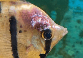
3. Piscinoodinium
Piscinoodinium is a sedentary flagellate that attaches to the skin, fin, and gills of fish. The common name for Piscinoodinium infection is Gold Dust or Velvet Disease.
The parasite has an amber pigment, visible on heavily infected fish. Affected fish will flash, go off feed, and die. Piscinoodinium is most pathogenic to young fish. The life cycle of this parasite can be completed in 10–14 days at 73–77°F, but lower temperatures can slow the life cycle.
Also, the cyst stage is highly resistant to chemical treatment. Therefore, several applications of treatment may be necessary to eliminate the parasite. For non-food species, chloroquine (10mg/L prolonged bath) has been reported to be efficacious.
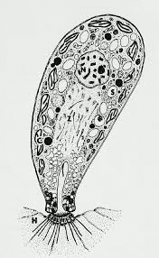
4. Cryptobia
Cryptobia is a flagellated protozoan common in cichlids. They are often mistaken for Hexamita as they are similar in appearance. However, Cryptobia are more drop-shaped, with two flagella, one on each end. Additionally, Cryptobia wiggles in a dart-like manner, whereas Hexamita spirals.
Cryptobia typically is associated with granulomas, in which the fish walls off the parasite. These parasites have been observed primarily in the stomach, but may be present in other organs. Fish afflicted with Cryptobia may become thin, lethargic and develop dark skin pigmentation.
A variety of treatments are presently being studied with limited success. Nutritional management has proven to take an active role in its control.
5. Myxozoa
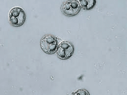
Myxozoa are parasites that are widely dispersed in native and pond- reared fish populations. Most infections in fish create minimal problems, but heavy infestations can become serious, especially in young fish.
Myxozoans are parasites affecting a wide range of tissues. They are an extremely abundant and diverse group of organisms, speculated by spore shape and size. Spores can be observed in squash preparations of the affected area at 200 or 400 x magnifications or by histologic sections.
Clinical signs vary, depending on the target organ. For example, fish may have excess mucus production, observed with Henneguya infections. This is a very large group of parasites which can cause disease in a wide variety of fishes.
They are obligate parasites of tissue (histozoic forms that reside in intercellular spaces or blood vessels that reside intracellularly) and organ cavities.
Key characteristics of the Myxozoa include development of a multicellular spore, presence of polar capsules in their spores and endogenous cell cleavage in both the trophozoite and sporogony stages.
The method of transmission of myxozoans is unknown, but evidence suggests that at least some pathogenic myxozoans have an indirect life cycle.
This life cycle may require the completion of two different life cycles involving a vertebrate (fish) and an invertebrate (annelid) host with each life cycle having its own sexual and asexual stages.
Severe infestations by these parasites can result in disease and/or death of the host fish. Each parasite is somewhat species specific as well as organ specific. A few of the more common myxozoan parasites are discussed below.
White or yellowish nodules may appear on target organs. Chronic wasting disease is common among intestinal myxozoans such as with Chloromyxum. Whirling disease caused by Myxoboluscerebralis has been a serious problem in salmonid culture.
Elimination of the affected fish and disinfection of the environment is the best control of myxozoans. There are no established remedies for fish. Spores can survive over a year, so disinfection is mandatory for eradication.
Read Also: Fish Biology: Anatomy, Physiology, Embryology and Endocrinology
6. Microsporidia
Microsporidians are intracellular parasites that require host tissue for reproduction. Fish acquire the parasite by ingesting infective spores from infected fish or food. Replication within spores (schizogony) causes enlargement of host cells (hypertrophy).
Infected fish may develop small tumor-like masses in various tissues. Diagnosis is confirmed by finding spores in affected tissues, either in wet mount preparations, or in histologic sections.
Clinical signs depend on the tissue infected and can range from no visible lesions to mortalities. In the most serious cases, cysts enlarge to a point that organ function is impaired and severe morbidity and/or mortality results. A common microsporidian infection is Pleistophora, which infects skeletal muscle.
There is no treatment for microsporidian infections in fish. Spores are highly resistant to environmental conditions and can survive for long periods. Elimination of the infected stock and disinfection of the environment is recommended.
7. Coccidia
Coccidia are intracellular parasites described in a variety of wild- caught and cultured fish. Their role in the disease process is poorly understood, but there is increasing evidence that they are potential pathogens. The most common species encountered in fish are intestinal infections.
Inflammation and death of the tissue can occur, which can affect organ function. Other infection sites include reproductive organs, liver, spleen, and swim bladder.
Clinical signs depend on target organ affected but may include general malaise, poor reproductive capacity, and chronic weight loss. A definitive diagnosis of tissue coccidia should be completed with histologic or electron microscopy.
Several compounds have been used to control coccidiosis with some success; however, consultation with an experienced fish health professional is recommended. Maintaining a proper environment and reducing stress appear to be important in preventing coccidia outbreaks in cultured fish.
In summary, flagellated protozoan infection of fish includes Hexamita or Spironucleus, Ichthyobodo, Piscinoodinium, Cryptobia, Myxozoa, Microsporidia, and Coccidia which affect freshwater fish and tropical fish . Elimination of the affected fish and disinfection of the environment is the best control.