This article explores fertilization and post-fertilization developments, including important phenomena in the life cycle of Angiosperms such as apomixis, adventive embryony, polyembryony, and parthenocarpy.
The ability to reproduce is one of the most essential characteristics of life, aimed at sustaining individual species. Reproduction in plants occurs in two main ways: asexual and sexual.
In flowering plants, sexual reproduction requires the fusion of two gametes one from the male organ and one from the female organ of the plant. This fusion results in the formation of a zygote, and the process is called fertilization.
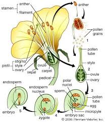
In Angiosperms, fertilization starts when compatible pollen (male gametophyte) reaches the stigma and concludes with the fusion of male and female gametes in the embryo sac (female gametophyte). The pollen, received by the gynoecium (female reproductive organ), is held at the stigma.
Pollen cannot directly reach the egg in the embryo sac. To solve this, the pollen germinates on the stigma and forms a pollen tube, which penetrates the stigmatic tissue, grows down the style, enters the ovary, and eventually reaches the embryo sac through the ovule. Once there, two sperm cells are released near the female gametes.
One sperm fuses with the egg (syngamy) to form the zygote, while the other fuses with the polar nuclei (triple fusion) to form the primary endosperm nucleus. This unique process, known as double fertilization, is characteristic of Angiosperms.
After multiple divisions, the primary endosperm nucleus develops into the endosperm, a highly nutritious tissue that nourishes the developing embryo. The zygote forms the embryo, which can be either dicotyledonous or monocotyledonous, depending on the plant species.
Read Also: All You Need To Know About Animal Shelters
Fertilization in Angiosperms

“Fertilization is the process of fusion between two dissimilar reproductive units called gametes.” In flowering plants, the process of fertilization was first described by Strasburger in 1884. As mentioned earlier, the female gametophyte (embryo sac) of Angiosperms is located in the ovule, far from the stigma.
Since the gynoecium (pistil) lacks any mechanism to directly transfer pollen from the stigma to the embryo sac, the pollen germinates on the stigma and produces a pollen tube to transport the male gametes to the embryo sac.
Stages of Fertilization in Angiosperms
1. Germination of Pollen Grains and Growth of Pollen Tube: When pollen is shed from the anther, it typically contains two cells: a generative cell and a vegetative (tube) cell. The generative cell forms two male gametes. Once the pollen lands on a receptive stigma through pollination, it hydrates, absorbing water and swelling.
The vegetative cell then forms a pollen tube. The stigmatic fluid contains sugars, lipids, and resins that provide a suitable medium for pollen germination.
Both the pollen grain and the pollen tube contain an enzyme called cutinase, which helps degrade the cutin on the stigma, allowing the pollen tube to penetrate the stigmatic tissue. The entire content of the pollen, including the two male gametes, moves into the pollen tube.
2. Growth of the Pollen Tube Through the Style: The pollen tube continues to grow through the stigmatic tissue, pushing its way through the style to reach the ovary. The style can be either hollow or solid. In a hollow style, the pollen tube grows along the epidermal surface, whereas, in a solid style, the pollen tube travels through the intercellular spaces between cells.
Pollen Tube Entry into the Ovule
After reaching the ovary, the pollen tube enters the ovule through one of three possible routes:
1. Through the Micropyle (Porogamy): This is the most common route, where the pollen tube enters through the micropyle, and the process is termed porogamy.
2. Through the Chalazal End (Chalazogamy): In this less common method, the pollen tube enters through the chalazal end of the ovule, seen in plants such as Casuarina, Betula, and Juglans regia.
3. Through the Integuments or Funiculus (Mesogamy): In mesogamy, the pollen tube enters through the integument (as in Cucurbita) or the funiculus (as in Pistacia).
Read Also: All You Need To Know About Sweet William Flowers
Entry of Pollen Tube into the Embryo Sac

Regardless of how the pollen tube enters the ovule, it always reaches the embryo sac through the micropylar end. This indicates that the pollen tube’s entry into the ovule does not affect its entry into the embryo sac. Once it passes through the micropyle, the pollen tube can follow different routes to enter the embryo sac:
- Between the egg cell and one of the synergids, as seen in Fagopyrum.
- Between the wall of the embryo sac and one of the synergids, as seen in Cardiospermum.
- Directly through one of the synergids, as seen in Oxalis.
Synergids play a crucial role not only in guiding the pollen tube into the embryo sac but also in the release of male gametes within the embryo sac.
Discharge of Male Gametes from the Pollen Tube
After the pollen tube reaches the embryo sac, it bursts at the tip, releasing the two male gametes. Before the tube bursts, the tube nucleus disintegrates. The male gametes then exhibit amoeboid movement: one moves toward the egg, and the other moves toward the polar nuclei.
Syngamy: Fusion of Gametes
When one of the male gametes reaches the egg, it fuses with it, forming a diploid zygote (2n) because both the egg and the male gamete are haploid. This fusion of male and female gametes is known as fertilization or syngamy. This phenomenon was first observed by Strasburger in 1884 in Monotropa.
The fate of the second male gamete was clarified by S.G. Nawaschin in 1898. While studying Fritillaria and Lilium, he discovered that the second male gamete fuses with the polar nuclei in a process called triple fusion. This discovery led to the concept of double fertilization, a hallmark of Angiosperms.
Post-Fertilization Developments
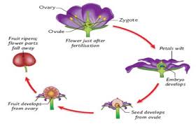
After fertilization, both the embryo and endosperm begin to develop within the ovule. The zygote, formed by the fusion of one male gamete with the egg, develops into the embryo.
Simultaneously, the primary endosperm nucleus, formed by triple fusion, develops into the endosperm. Other cells within the embryo sac, such as synergids and antipodal cells, disorganize after fulfilling their role.
Development of the Endosperm

The primary endosperm nucleus is the product of triple fusion. It undergoes several divisions to form the endosperm, which is a triploid (3n) tissue unique to Angiosperms.
The endosperm provides essential nutrients to the developing embryo. It is distinct from the haploid endosperm found in heterosporous Pteridophytes and Gymnosperms, where no triple fusion occurs.
Three types of endosperm development have been identified:
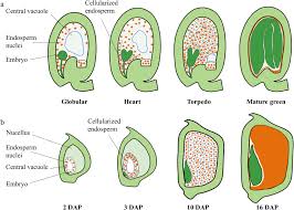
1. Nuclear Endosperm: In this type, the division of the primary endosperm nucleus is not followed by immediate wall formation. The resulting nuclei remain free within the cytoplasm of the embryo sac, forming a peripheral layer around a large central vacuole.
Wall formation occurs later, usually starting from the periphery and moving inward, as seen in Arachis hypogaea. In some cases, such as in palms (Cocos nucifera), the central vacuole may remain even in mature seeds. The liquid endosperm in coconuts, known as coconut milk, contains growth-promoting factors and is commonly used in plant tissue culture.
2. Cellular Endosperm: In this type, the division of the primary endosperm nucleus is immediately followed by wall formation, resulting in a cellular structure from the outset. The first wall is laid transversely, but subsequent divisions occur irregularly. Examples include Adoxa, Peperomia, and Villarsia.
3. Helobial Endosperm: This type is found in members of Helobiales, such as Vallisneria and Eremurus. It represents an intermediate form between nuclear and cellular endosperms. The first division of the primary endosperm nucleus is followed by the formation of a transverse wall, dividing the embryo sac into a small chalazal chamber and a large micropylar chamber.
Subsequent free nuclear divisions occur in both chambers, but the micropylar chamber undergoes more divisions. Eventually, the chalazal chamber degenerates, and the micropylar chamber forms a cellular endosperm.
The development of the embryo follows the fertilization process in plants, where changes in the ovule result in seed formation. After a period of rest, the oospore (zygote) undergoes embryogenesis, which leads to the mature embryo.
Though monocotyledons and dicotyledons start embryo development similarly, they later differ due to the number of cotyledons.
1. Dicotyledonous Embryo: It has two cotyledons attached to the embryonic axis.
2. Monocotyledonous Embryo: It has a single cotyledon at the apex of the embryonic axis.
In all angiosperms, embryogenesis begins with the division of the oospore into a two-celled proembryo, with the basal cell forming the suspensor and the terminal cell (embryo cell) responsible for further embryo development.
Apomixis
Apomixis, meaning reproduction without fertilization, involves the asexual formation of seeds without the processes of meiosis or syngamy. This process was discovered in Citrus seeds and results in offspring that are genetically identical to the female parent, known as apomictic seeds.
Apomixis is common in Poaceae, Asteraceae, and other families. It can be obligate (only method of reproduction) or facultative (both gametic and apomictic reproduction occur).
Types of apomixis include:
1. Non-recurrent apomixis: Haploid parthenogenesis or haploid apogamy produces sterile embryos.
2. Recurrent apomixis: Diploid parthenogenesis/apogamy results in viable embryos.
3. Adventive apomixis: Embryos develop from somatic cells outside the embryo sac.
4. Vegetative apomixis: Reproduction occurs through vegetative buds or bulbils instead of seeds.
Parthenogenesis
Parthenogenesis is the development of an embryo directly from an unfertilized egg, leading to haploid or diploid embryos. It can be:
1. Haploid parthenogenesis: The embryo and resulting plant are haploid and sterile.
2. Diploid parthenogenesis: A diploid egg develops into a diploid embryo.
Polyembryony
Polyembryony refers to the occurrence of multiple embryos within a single seed. It is more common in gymnosperms but can also occur in angiosperms like Citrus species. There are two main types:
1. True polyembryony: Multiple embryos develop within the same embryo sac.
2. False polyembryony: Embryos develop in separate sacs within the same ovule.
Parthenocarpy
Parthenocarpy is the development of fruits without fertilization, leading to seedless fruits. This phenomenon can be:
1. Stimulative parthenocarpy: Requires pollination stimulus.
2. Vegetative parthenocarpy: Occurs in unpollinated flowers.
Parthenocarpy can also be induced:
1. Genetic: Through mutations or hybridization (e.g., seedless oranges).
2. Environmental: Due to environmental stress like frost or temperature fluctuations.
3. Chemically induced: Using plant growth regulators like auxins and gibberellins to produce seedless fruits.
Do you have any questions, suggestions, or contributions? If so, please feel free to use the comment box below to share your thoughts. We also encourage you to kindly share this information with others who might benefit from it. Since we can’t reach everyone at once, we truly appreciate your help in spreading the word. Thank you so much for your support and for sharing!

