The biology of fishes is the biology of active living organisms in water. It introduces learners to the basics of fish biology, which entails: Anatomy, Physiology, Embryology and Endocrinology of bony and cartilaginous fish.
Fishes are cold blooded or poikilothermic animals i.e. their body temperature varying passively in accordance with the ambient temperature (surrounding water temperature).
Although, fishes as a group can tolerate wide range of temperature just below 00C to 450C, individual species generally have a preferred or optimum as well as a more restricted temperature range.
Procedure – Activity
Fish dissection to reveal the internal anatomical features of fish in the three living groups of fishes (cyclostomes, chondrichthyes and the osteichthyes. Demonstration of respiration, circulation or skeletal system in fish using plastic models.

1. External Fish Anatomy
At your lab stations, you will have a dissection kit. Please be careful with the scalpel as they are very sharp. Bring the pan to the front bench and get a fish.
Back at your station, and using the descriptions below, identify the external structures of your fish writing the answers to any questions that are posed.
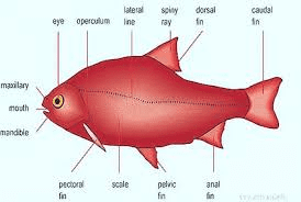
1. Eyes – Fish eyes serve a variety of purposes – to seek out food, to avoid predators and other dangers, and, perhaps even to navigate in the ocean. Fish do not have eyelids.
They are constantly bathed in water and do not need tears. Using your finger, gently move the eye in its socket. Is there an eyelid present?

2. Nostrils – Some fish have a well-developed sense of smell and use this ability to seek out their home streams for spawning. In some cases, this scent is also helpful in avoiding predators. Fish breathe through their gills, not their nostrils.
3. Lateral Line – Fish do not have ears, as such. In part, low frequency sounds are detected in the water through a system of small holes along each side of a fish called the lateral line, which is connected to a delicate system of nerves. They also react to medium frequencies suggesting they detect these as well.
4. Mouth – Fish use their mouth to catch and hold food of various types, but their food is not chewed before swallowing, it is swallowed whole. The mouth is the beginning of the fish’s alimentary canal (digestive tract).
In addition, it is a very important part of the breathing process. Water is constantly taken in through the mouth and forced out over the gills where oxygen is extracted. Inside the mouth for teeth. Open and close the mouth. Describe how the upper and lower jaw articulates during this movement.
Examine the upper and lower jaw. Does the lower jaw stick out further? This would mean the fish eats by attacking its prey from below. Or does the upper jaw stick out further? This would mean the fish eats by attacking its prey from above.
Do both jaws meet at one common point? This fish eats by attacking from above or below. What direction do you think your fish attacks its prey from and why?
5. Vent – The external opening of the alimentary canal. Urine, feces, eggs and milt exit here.
6. Gills – Fish gills are composed of two basic parts, the gill covers and the gill filaments. The gill cover, a bony structure called the operculum, protects delicate filaments and, together with the mouth, forces water containing oxygen over the gills. The gills are probably one of the most important organs in the body of a fish. They are delicate but very effective breathing mechanisms.
Gills are far more efficient than human lungs, because they extract 80% of the oxygen dissolved in water, while human lungs only extract 25% of the oxygen in the air.
Gills are thin walled structures, filled with blood vessels. Their structure is arranged so that they are constantly bathed in water. The fish takes in the water through its mouth.
The oxygen dissolved in the water is absorbed through the thin membranes into the fish’s blood. Carbon dioxide is simultaneously released from the blood into the water across the same membranes.
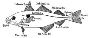
7. Fins – Fish have two sets of paired fins (pelvic and pectoral) and four single fins (dorsal, caudal and anal). Fish can contract their muscles and move the pelvic and pectoral fins for movement in all directions.
The caudal fin is used for forward momentum. The dorsal and anal fins aid in stabilizing the fish in the water and preventing it from rolling. All fish fins are made of bony fin rays that are connected to each other with a thin membranous tissue.
2. Internal Fish Anatomy

Place the fish on its side in the dissection pan, belly towards you, head pointing to your right. Insert a pair of sharp dissection scissors into the vent and make a shallow cut up to and between the pectoral fins all the way to where the opercula meet.
Read Also: Introduction to Fishing Gear Technology
Locate the heart, it will be in the cavity anterior to the pectoral fins. Use the scissors to snip the aorta (large, white tube on top of the heart) and remove the heart.
The large, brownish organ in the body cavity posterior to the pectoral fins is the liver. It is used to synthesize and secrete the essential nutrients that were contained in the food.
It plays a part in maintaining the proper levels of blood chemicals and sugars. The gall bladder, which is attached to the liver, contains green bile which in part is used to help digest fats.
Locate and remove the alimentary canal. It starts at the esophagus which is connected to the mouth and ends at the intestines at the vent. Once removed, locate the following: Esophagus: muscular tube that moves food from the mouth to the stomach;
1. Stomach: a saclike organ that receives the food from the esophagus; mechanical digestion occurs here.
2. Intestines: tube running from the vent to the stomach; chemical digestion and nutrient absorption occurs here.
The air bladder is the only remaining organ in the body cavity. It is a whitish organ and the fish use it to control their buoyancy. They can inflate or deflate it with gas. Remove the air bladder.
The dark red line along the backbone is the kidney. The forward part of the kidney of a fish functions to replace red blood cells, the rearward part filters waste out of the blood. The kidney can be removed by slicing through the membrane along each side, and then scraping with a spoon.
What is left is the body cavity, or coelom, that houses major organs. If your fish is female, you should find the ovaries near the vent, they are an orange mass of eggs. Fish lay thousands of eggs and only a small percentage ever makes it to adulthood. If your fish is male, you should find a bladder of milt, or fish sperm, near the vent.
Reproduction is carried out when the female deposits her eggs into the water and the male quickly fertilizes them with his sperm, this is called external fertilization. Any resulting fertilized eggs will develop in the water column without aid from the parents.
Endocrine System
The endocrine system is made up of specialized cells, glands and hormones. Acting like a communication network, it responds to stimuli by releasing hormones, the chemical messengers that carry instructions to target cells throughout the body, from endocrine glands.
In summary, the gross external anatomy allows an individual especially the fisheries scientist to identify most species with a fair degree of accuracy.
All vertebrate animals such as fish, amphibians, reptiles, birds and mammals, including humans have the same general endocrine glands and release similar hormones to control development, growth, reproduction and other responses.
Read Also: Introduction to Fish Nutrition
Read Also: Impact of Agricultural Wastes on Human and Environment
