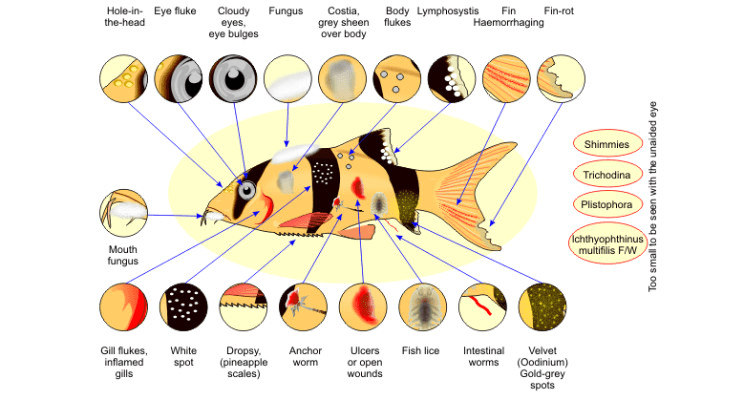
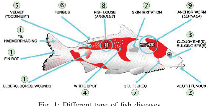
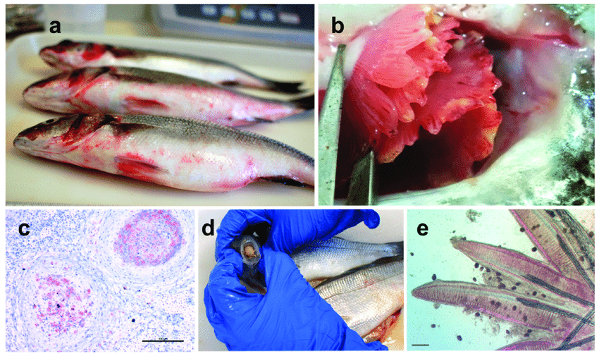
A common mistake of fish culturists is misdiagnosing disease problems and treatment of fish with the wrong medication or chemical. When the chemical doesn’t work, they will try another, then another.
Selecting the wrong treatment because of misdiagnosis is a waste of time and money and maybe more detrimental to the fish than no treatment at all. The majority of fish parasites can only be identified by the use of a microscope.
If a microscope is unavailable, or the person using it has no previous experience with one, the diagnosis is difficult and questionable. Successful fish culturists learn by experience.
New comers to the field need to learn the fundamentals of diagnostic procedures and how to use a microscope to identify parasites by attending short training courses.
The following descriptions of common parasites can be used as references for understanding a professional diagnostic report or as a quick reference for the experienced fish culturist.
Protozoa

Most of the commonly encountered fish parasites are protozoan. With practice, these can be among the easiest to identify and are usually among the easiest to control.
The protozoan is single-celled organism, many of which are free-living in the aquatic environment. Typically, no intermediate host is required for the parasite to reproduce (direct life cycle).
Consequently, they can build up to very high numbers when fish are crowded causing weight loss, debilitation, and mortality. Five groups of protozoans are described below: ciliates, flagellates, myxozoans, microsporidians, and coccidia.
Parasitic protozoan in the latter three groups can be difficult or impossible to control as discussed below.

1. Ciliates
Most of the protozoans identified by aquarists will be ciliates. These organisms have tiny hair-like structures called cilia that are used for locomotion and/or feeding.
Ciliates have a direct life cycle and many are common inhabitants of pond-reared fish. Most species do not seem to bother host fish until numbers become excessive. In aquaria, tanks, and ponds which are usually closed systems, ciliates should be eliminated.
Uncontrollable or recurrent infestations with ciliated protozoan are indicative of a husbandry problem. Many of the parasites proliferate in organic debris accumulated in the bottom of a tank or vat.
Ciliates are easily transmitted from tank to tank by nets, hoses, or caretakers’ wet hands. Symptoms typical of ciliates include skin and gill irritation displayed by flashing, rubbing, and rapid breathing.
2. Ichthyophthirius multifiliis
The disease called Ich or white spot disease has been a problem to aquarists for generations. Fish infected with this organism typically develop small blister-like raised lesions along the body wall and/or fins.
If the infection is restricted to the gills, no white spots will be seen. The gills will appear swollen and be covered with thick mucus. Identification of the parasite on the gills, skin, and/or fins is necessary to conclude that fish has an ich infection.
The mature parasite is very large, up to 1000 µm in diameter, is very dark in color due to the thick cilia covering the entire cell, and moves with an amoeboid motion. Classically, I. multifiliis is identified by its large horseshoe-shaped macronucleus.
This feature is not always readily visible, however, and should not be the sole criterion for identification. Immature forms of I. multifiliis are smaller and more translucent in appearance.
Some individuals have suggested that the immature forms of I. multifiliis resemble Tetrahymena. Fortunately, scanning the preparation will usually reveal the presence of mature parasites and allow confirmation of the diagnosis.
If only one parasite is seen, the entire system should be treated immediately. Ich is an obligate parasite and is capable of causing massive mortality within a short time. Because the encysted stage is resistant to chemicals, a single treatment is not sufficient to treat Ich.
Repeating the selected treatment every other day at water temperatures 68–77°F for three to five treatments will disrupt the life cycle and control the outbreak. Daily cleaning of the tank or vat helps to remove encysted forms from the environment
Read Also: Introduction to Fisheries Ecology

3. Chilodonella
Chilodonella is a ciliated protozoan that causes infected fish to secrete excessive mucus. Infected fish may flash and show similar signs of irritation. Many fish die when infestations become moderate which includes five to nine organisms per low power field on the microscope to heavy which is greater than ten organisms per low power field.
Chilodonella is easily identified using a light microscope to examine scrapings of skin mucus or gill filaments. It is a large, heart-shaped ciliate (between 60 to 80m) with bands of cilia along the long axis of the organism. The organism is easily recognized at 100X magnification.
Chilodonella can is controlled with any of the chemicals listed in Table 1, and one treatment is usually adequate. Chilodonella has have been eliminated in tanks using recirculating water systems by maintaining 0.02% salt solution.
4. Tetrahymena
Tetrahymena is a protozoan commonly found living in organic debris at the bottom of an aquarium or tank. Tetrahymena is a teardrop-shaped ciliate that moves along the outside of the host.
The presence of Tetrahymena on the body surface in low numbers (less than five organisms per low power field) is probably not significant. It is commonly found on dead material and is associated with high organic loads.
Therefore, observing Tetrahymena on fish, which have been on the tank bottom, does not imply the parasite is the primary cause of death.
Identification of Tetrahymena internally is a significant but untreatable problem. A common site of internal infection is the eye. Affected fish will have one or both eyes markedly enlarged (exophthalmia).
Squash preparations made from fresh material reveal large numbers (≥ 10 per low power field) of Tetrahymena associated with fluids in the eye. Fish infected with Tetrahymena internally should be removed from the collection and destroyed.
5. Trichodina
Trichodina is one of the most common ciliates present on the skin and gills of pond-reared fish. Low numbers which involves less than five organisms per low power field are not harmful, but when fish are crowded or stressed, and water quality deteriorates, the parasite multiplies rapidly and causes serious damage.
Typically, heavily infested fish do not eat well and lose condition. Weakened fish become susceptible to opportunistic bacterial pathogens in the water.
Trichodina can be observed on scrapings of skin mucus, fin, or on gill filaments. Its erratic darting movement and the presence of a circular, toothed disc within its body easily identify it. One application should be sufficient. Correction of environmental problems is necessary for complete control.
Read Also: The External Anatomy of Cartilaginous Fish
6. Ambiphyra
Ambiphyra, previously called Scyphidia, is a sedentary ciliate that is found on the skin, fins, or gills of host fish. Its cylindrical shape, row of oral cilia, and middle bank of cilia identify Ambiphyra.
It is common on pond-reared fish, and when present in low numbers (less than five organisms per low power field), it is not a problem. High organic loads and deterioration of water quality are often associated with heavy, debilitating Ambiphyra infestations.
7. Apiosoma
Apiosoma, formerly known as Glossatella, is another sedentary ciliate common on pond-reared fish. Apiosoma can cause disease if their numbers become excessive. The organism can be found on gills, skin, or fins. The vase-like shape and oral cilia are characteristic.
8. Epistylis
Epistylis is a stalked ciliate that attaches to the skin or fins of the host. Epistylis is of greater concern than many of the ciliates because it is believed to secrete proteolytic which means protein-eating enzymes that create a wound, suitable for bacterial invasion, at the attachment site. It is similar in appearance to Apiosoma except for the non-contractile long stalk and its ability to form colonies.
In contrast to the other ciliates discussed above, the preferred treatment for Epistylis is salt. Fish can be placed into a 0.02% salt solution as an indefinite bath, or a 3% salt dip. More than one treatment may be required to control the problem.
9. Capriniana
Capriniana, historically called Trichophyra, is a sessile ciliate that attaches to the host’s gills with a sucker. They have characteristic cilia attached to an amorphous-shaped body. In heavy infestations, Capriniana can cause respiratory distress in the host.
Read Also: Impact of Agricultural Wastes on Human and Environment
