This article gives a basic knowledge of what is meant by digestion, the importance of digestion, how food is digested, absorption and transport of nutrients, and how the digestive process is controlled. The involvement of hormones and nerve regulators will also be discussed.
Understanding Anatomy in Farm Animals
Anatomy is the science of the morphology or structure of animals or plants. It involves the dissecting of an animal or plant in order to determine the position, structure, alignment etc. of its parts. The structure of an organism or body, or a model of it as dissected is also its anatomy.
Meaning of Physiology in Animal Body Function
Physiology is the branch of biology involved with or dealing with the functions and vital processes of living organisms or their parts and organs or systems.
Definition of Domestic Animals in Agriculture
This refers to animals that have been tamed, such that they are kept under the care of man.
Feeding Habits and Dentition of Farm Animals
The dentition of animals suits their feeding habits.
The dental formula of three typical herbivores is as follows:
1. Rabbit
I 2/1
C 0/0
PM 3/2
M 3/3
= 28
2. Horse
I 3/3
C 1/1
PM 3/3
M 3/3
= 40
3. Sheep
I 0/3
C 0/1
PM 3/3
M 3/3
= 32
In a mammal, the teeth or dental formula indicate quite clearly the feeding habits of their owner. For man, the teeth of mammals fall into four categories: In the front of the mouth are the chisel-shaped incisors (I) which are followed by a single sharp canine.
Behind this are the premolars (PM) which may be modified for grinding but do not usually show the complex cusp patterns of the back teeth or molars (M).
In herbivores, the incisors are large and are continuously self-sharpening by having a leading edge of enamel working against softer dentine in the opposing tooth. The canines are usually absent and in their place a large gap, or diastema, exists which allows the food to be freely circulated in the mouth.
The premolars tend to become molarform and work in conjunction with the molars, the two types of teeth making up a broad grinding surface. Like the incisors, they show continuous growth and also the most specialized of mammalian cusp patterns.
In carnivores, all the teeth find some use in feeding and, on the whole, there tend to be more of them than in the herbivores. In the front of the mouth, the incisors are not particularly well developed though they find some use in grooming and nibbling meat from bones.
Behind the incisors are a pair of very large canines in each jaw and these stabbing teeth are used to catch prey. The premolars and molars are covered in enamel and rise to sharp edges along the line of the jaw, the joint action of upper and lower teeth providing a powerful shearing action like scissors.
These teeth, adapted for cutting and bone crunching, are called the carnassials and are found in the cat family and other families of carnivores.
Read Also : How to Plant Fruit Trees for Optimum Performance
Stomach Structures in Ruminant and Non-Ruminant Farm Animals

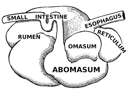
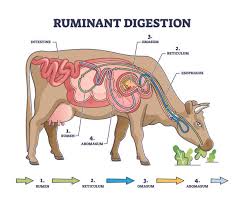
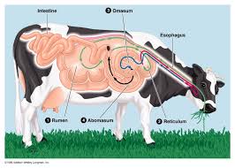
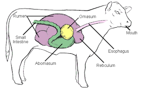
Animals have varying structures and functions of the stomach depending on the type of farm animal. Cattle, sheep, and goats have stomach with compartments. Such animals have a large pouch known as the rumen.
This is virtually a storage compartment in which newly eaten but un-chewed food is passed. Some digestion of cellulose and vitamin D synthesis occur due to bacterial action taking place in the rumen.
Some of the products of cellulose digestion are absorbed by the rumen. From the rumen, food passes to a smaller compartment called the reticulum. The food is then regurgitated by anti-peristaltic movement into the mouth where it is masticated.
The food in form of pulp is again swallowed and moved to the third (3rd) compartment known as omasum, and from here is then passed into the true stomach which is the fourth (4th) compartment called the abomasum. Animals with this complicated stomach structure (as in Fig. 1) are known as ruminants.
Monogastric and Pseudo-Ruminant Digestive Systems
A monogastric animal has a simple single-chambered stomach, compared to the ruminants. This includes almost all mammals, except for ruminants like cows and oxen. There are various species of monogastric animals ranging from humans, cats, dogs, mice, elephants, pigs etc.
The difference is that monogastric – simple stomach i.e., just a bag. Ruminant – stomach has several compartments. Ruminants also regurgitate their food to chew on it further.
A pseudo-ruminant is the classification of an animal based on its digestive tract. These types of animals are still considered foregut fermenters, but only have three chambers in their stomach, not four like true ruminants do. Pseudo means ‘false’ so they are ‘false’ ruminants.
The chambers are basically the reticulum, omasum and abomasum. They do not have the characteristic rumen that identifies ruminants; instead, they have an enlarged cecum. The animals that are often referred to as pseudo-ruminants are all camelids (camels, alpacas, llamas, etc).
The Digestive System of Farm Animals

The digestive system is made up of the digestive tract – a series of hollow organs joined in a long, twisting tube from the mouth to the anus – and other organs that help the body break down and absorb food.
Organs that make up the digestive tract are the mouth, oesophagus, stomach, small intestine, large intestine – also called the colon – rectum, and anus. Inside these hollow organs is a lining called the mucosa.
In the mouth, stomach, and small intestine, the mucosa contains tiny glands that produce juice to help digest food. The digestive tract also contains a layer of smooth muscle that helps break down food and move it along the tract.
Two digestive organs, the liver and the pancreas, produce digestive juices that reach the intestine through small tubes called ducts. The gallbladder stores the liver’s digestive juices until they are needed in the intestine. Parts of the nervous and circulatory systems also play major roles in the digestive system.
Importance of Digestion in Animal Body Function
When food such as bread, meat, and vegetables is eaten, it is not in a form that the body can use as nourishment. Food and drink must be changed into smaller molecules before they can be absorbed into the blood and carried to cells throughout the body.
Digestion is the process by which food and drink are broken down into their smaller parts so the body can use them to build and nourish cells and to provide energy.
How Food is Digested in Animals
Digestion involves mixing food with digestive juices, moving it through the digestive tract, and breaking down large molecules of food into smaller molecules. Digestion begins in the mouth, when food is chewed and swallowed, and is completed in the small intestine.
Movement of Food through the Digestive Tract
The large, hollow organs of the digestive tract contain a layer of muscle that enables their walls to move. The movement of organ walls can propel food and liquid through the system and can also mix the contents within each organ.
Food moves from one organ to the next through muscle action called peristalsis. Peristalsis looks like an ocean wave traveling through the muscle.
The muscle of the organ contracts to create a narrowing and then propels the narrowed portion slowly down the length of the organ. These waves of narrowing push the food and fluid in front of them through each hollow opening.
The first major muscle movement occurs when food or liquid is swallowed. Although swallowing can be started by choice, once the bolus touches the throat it becomes involuntary and proceeds under the control of the nerves.
Swallowed food is pushed into the esophagus, which connects the throat above with the stomach below. At the junction of the esophagus and stomach there is a ring-like muscle, called the lower esophageal sphincter, closing the passage between the two organs.
As food approaches the closed sphincter, the sphincter relaxes and allows the food to pass through to the stomach.
The stomach has three mechanical tasks. First, it stores the swallowed food and liquid. To do this, the muscle of the upper part of the stomach relatively accepts large volumes of swallowed material.
The second job is to mix up the food, liquid, and digestive juice produced by the stomach. The lower part of the stomach mixes these materials by its muscle action. The third task of the stomach is to empty its contents slowly into the small intestine.
Several factors affect emptying of the stomach, including the kind of food and the degree of muscle action of the emptying stomach and the small intestine. Carbohydrates, for example, spend the least amount of time in the stomach, while protein stays in the stomach longer, and fats the longest.
The food dissolves into the juices from the pancreas, liver, and intestine. The contents of the intestine are mixed and pushed forward to allow further digestion.
The digested nutrients are absorbed through the intestinal walls and transported throughout the body. The waste products of this process are undigested parts of the food, fibre, and older cells that have been shed from the mucosa. These materials are pushed into the colon, where they remain until the faeces are expelled by a bowel movement.
Read Also: 12 Medicinal Health Benefits Of Renealmia alpinia (Pink cone ginger)
Digestion of Food in Domestic Animals

The initial digestive glands involved are the salivary glands located in the mouth. Saliva produced by these glands contains an enzyme that initiates the breakdown of starch into smaller molecules. An enzyme is a substance that accelerates chemical reactions within the body.
Subsequently, the stomach lining houses the next set of digestive glands. These glands secrete stomach acid and an enzyme responsible for protein digestion. A thick mucus layer coats the mucosa, preventing the acidic digestive juice from damaging the stomach tissue. Typically, the stomach mucosa effectively releases the juice without harming the body’s tissues.
Upon the stomach emptying its contents (chyme) into the small intestine, digestive juices from two additional organs mix with the food. The pancreas produces a juice rich in enzymes that decompose carbohydrates, fats, and proteins. Additional enzymes active in this process originate from glands in the intestinal wall.
The liver, another vital organ, produces bile—a digestive juice stored in the gallbladder between meals. During meals, bile is released into the intestine via bile ducts to emulsify fats, facilitating their digestion by pancreatic and intestinal enzymes.
Absorption and Transport of Nutrients
The majority of digested food molecules, along with water and minerals, are absorbed through the small intestine. The mucosa of the small intestine features numerous folds adorned with tiny fingerlike projections called villi, which are further covered with microvilli.
These structures significantly increase the surface area for nutrient absorption. Specialized cells enable absorbed materials to traverse the mucosa into the bloodstream, distributing them throughout the body for storage or further chemical transformation. Water absorption primarily occurs in the large intestine.
1. Carbohydrates: Dietary guidelines recommend that 45 to 65 percent of total daily calories derive from carbohydrates. Foods abundant in carbohydrates include bread, potatoes, dried peas and beans, rice, pasta, fruits, and vegetables, many of which contain both starch and fiber.
Digestible carbohydrates starch and sugars are decomposed into simple molecules by enzymes present in saliva, pancreatic juice, and the small intestine lining.
Starch digestion involves two steps: initially, an enzyme in saliva and pancreatic juice breaks down starch into maltose molecules; subsequently, an enzyme in the small intestine lining splits maltose into glucose molecules, which are absorbed into the bloodstream.
Glucose is transported to the liver for storage or utilized to supply energy for bodily functions.
An enzyme in the small intestine lining also digests sucrose (table sugar) into glucose and fructose, which are absorbed into the bloodstream. Milk contains lactose, another sugar transformed into absorbable molecules by an enzyme in the intestinal lining.
2. Fiber: Ruminants primarily rely on fiber sources for nutrients, whereas fiber digestion in pseudo-ruminants ranges from 40 to 70 percent, depending on the fiber type.
3. Protein: Foods such as meat, eggs, and beans comprise large protein molecules requiring enzymatic digestion before utilization in building and repairing body tissues.
An enzyme in stomach juice initiates protein digestion. In the small intestine, enzymes from pancreatic juice and the intestinal lining complete the breakdown of proteins into amino acids.
These amino acids are absorbed into the bloodstream and transported throughout the body to construct cellular structures.
4. Fats: Fat molecules serve as a concentrated energy source. The initial step in fat digestion involves emulsification into the intestine’s watery contents. Bile acids produced by the liver emulsify fats into tiny droplets, allowing pancreatic and intestinal enzymes to further break down large fat molecules into smaller ones, such as fatty acids and cholesterol.
Bile acids assist in transporting these molecules into mucosal cells, where they are reassembled into larger molecules, packaged into lymphatic vessels near the intestine, and eventually carried by the bloodstream to various body parts for storage.
5. Vitamins: Vitamins, essential dietary components, are absorbed through the small intestine. They are categorized based on solubility: water-soluble (B vitamins and vitamin C) and fat-soluble (vitamins A, D, E, and K). Fat-soluble vitamins are stored in the liver and fatty tissues, while excess water-soluble vitamins are excreted in urine.
6. Water and Salt: A significant portion of material absorbed through the small intestine consists of water and dissolved salts, originating from consumed food, liquids, and digestive gland secretions.
Control of The Digestive System
The digestive system’s functions are regulated by hormones produced and released by cells in the stomach and small intestine mucosa. These hormones enter the bloodstream, travel through the circulatory system, and return to the digestive organs to stimulate digestive juice secretion and organ movement.
Key hormones include:
1. Gastrin: Stimulates the stomach to produce acid for food digestion and supports normal cell growth in the stomach, small intestine, and colon linings.
2. Secretin: Prompts the pancreas to release bicarbonate-rich digestive juice, neutralizing stomach acid in the small intestine. It also stimulates pepsin production in the stomach and bile production in the liver.
3. Cholecystokinin (CCK): Encourages the pancreas to produce digestive enzymes and causes the gallbladder to release bile. It also supports normal pancreatic cell growth.
Additional hormones regulating appetite:
4. Ghrelin: Produced in the stomach and upper intestine when the digestive system is empty, stimulating appetite.
5. Peptide YY: Produced in response to food intake, suppressing appetite.
These hormones interact with the brain to regulate food intake based on nutrient needs.
Neural Regulation
Two nerve types control digestive system actions:
1. Extrinsic nerves: Originate from the brain or spinal cord, releasing chemicals like acetylcholine and adrenaline. Acetylcholine increases muscle contractions in digestive organs, enhancing food and juice movement. Adrenaline reduces blood flow to these organs, slowing digestion.
2. Intrinsic nerves: Located within the walls of the esophagus, stomach, small intestine, and colon. They respond to food-induced stretching of organ walls by releasing substances that adjust food movement and digestive juice production.
Together, nerves, hormones, blood, and digestive organs coordinate the complex processes of digesting and absorbing nutrients from consumed foods and liquids.
Do you have any questions, suggestions, or contributions? If so, please feel free to use the comment box below to share your thoughts. We also encourage you to kindly share this information with others who might benefit from it. Since we can’t reach everyone at once, we truly appreciate your help in spreading the word. Thank you so much for your support and for sharing!

