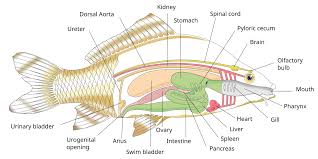
Good taxonomy is essential for ecological, biogeographical, and evolutionary studies of any group of organisms. Fish serve as hosts to a range of parasites that are taxonomically diverse and exhibit a wide variety of life cycle strategies.
Whereas many of these parasites are passed directly between ultimate hosts, others need to navigate through a series of intermediate hosts before reaching a host in (or on) which they can attain sexual maturity.
Read Also: 22 Medicinal Health Benefits of Nutmeg (Myristica Fragrans)
Trichodinids: Morphology and Taxonomy in Fish Health
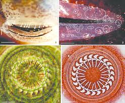
Trichodinids are members of the peritrichous ciliates, a paraphyletic group within the Oligohymenophorea. Specifically, they are mobiline peritrichs because they are capable of locomotion, as opposed to sessiline peritrichs such as Vorticella and Epistylis, which adhere to the substrate via a stalk or lorica.
There are over 150 species in the genus Trichodina. Trichodinella, Tripartiella, Hemitrichodina, Paratrichodina, and Vauchomia are similar genera. Trichodinids are round ciliates that may be disc-shaped or hemispherical.
The cytostome (cell mouth) is on the surface that faces away from the host; this is termed the oral surface. The other side, or adoral surface, attaches to the skin of the host or other substrate.
There is a spiral of cilia leading towards the cytostome and several rings of cilia at the periphery of the cell, responsible for creating adhesive suction and locomotory power.
In the taxonomy of trichodinids, the exact number, shape, and arrangement of the cytoskeletal denticles are critical for determining taxonomic relationships. These characters are usually revealed by silver nitrate staining of microscope slides, which stains the cell cytoplasm black and leaves the denticles white.
Cestoda: Morphology and Taxonomy in Fish Health
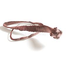
Cestodes are a taxonomic class of organisms in which the adult stage usually lives in the intestinal tract of vertebrates. Intermediate stages live in a wide variety of body locations in both vertebrate and invertebrate hosts.
The bodies of most cestodes are ribbon-shaped and divided into short segments called proglottids, hence the name “tapeworm”. Diagnosis of cestodiasis is dependent upon demonstration of the parasite within the intestinal tract of the fish.
Clinical signs of cestodiasis include emaciation, anemia, discoloration of the skin, and susceptibility to secondary infections. Low numbers of pleurocercoids may be located in vital organs such as the brain, heart, spleen, kidney, or gonad and have a devastating effect on the fish.
Cestodes, more commonly known as tapeworms, are entirely parasitic and can be found as adults all over the world inhabiting the intestines of their hosts. They are dorso-ventrally flattened, can reach lengths of several meters, and normally change hosts at least once in their life cycle.
The scolex or ‘head’ of the worm, which penetrates the intestine wall, is the most distinctive part of the parasite and is often used in determining species. Cestodes possess no gut, and nutritional uptake is mediated completely by the tegument, which is resistant to attack by the host’s digestive enzymes.
Larval tapeworms can be found in other organs of intermediate hosts, with transmission to the final host normally via the food chain.
Species of Cestodes Found in Freshwater Fish
Species found in freshwater fish include: Archigetes sieboldi, Bathybothrium rectangulum, Biacetabulum appendiculatum, Bothriocephalus acheilognathi, B. claviceps, Caryophyllaeides fennica, Caryophyllaeus fimbriceps, C. laticeps, Cyathocephalus truncates, Diphyllobothrium dendriticum, D. ditremum, D. latum, D. vogeli, Eubothrium crassum, E. fragilis, E. salvelini, Hepatoxylon squali, Khawia sinensis, Ligula intestinalis, Monobothrium wageneri, Proteocephalus ambiguous, P. cernua, P. exiguous, P. filicollis, P. macrocephalus, P. neglectus, P. osculates, P. parallacticus, P. percae, P. pollanicola, P. sagittus, P. torulosus, P. ocellatus, Schistocephalus solidus, S. pungitii, Scolex pleuronectis, Triaenophorus lucii, T. nodulosus, and Valipora campylancristrota.
Triaenophorus nodulosus: Classification and Morphology
1. Classification:
Kingdom: Animalia
Phylum: Platyhelminthes
Class: Cestoda
Order: Pseudophyllidea
Family: Triaenophoridae
Genus: Triaenophorus
Species: T. nodulosus
2. Size Range: Sexually mature adult worms can range from 65-380mm in length and 2-6mm in width. Eggs range from 0.052-0.071mm (average 0.063mm) in length and 0.033-0.045mm (average 0.042mm) in width.
3. Life Stage: Egg/Coracidium: Free-living environment: On the substrate or free-floating in the water column in shallow zones of fresh waters. Duration of stage: Embryonal development in the eggs and the hatching of the coracidia takes between 4 and 7 days at 17-20°C, with the coracidium fully formed by day 5.
The optimal temperature for development is 20°C. At 18-20°C, the coracidium can survive in its free-swimming state for 1-3 days. At lower temperatures, the coracidium seems to survive longer; 4 days at 15-16°C and up to 10-13 days at 2.5°C but will survive for less than an hour at 29°C.
4. Morphology: Originally, the coracidium is enclosed in a broad ciliated embryonal membrane, occupies a large amount of space in the egg, and moves actively. Recently hatched coracidia are 45-50µm x 44µm, while ones that are two or three days old are 100µm x 88µm, with an average size of 64.2µm x 58.9µm.
The coracidium has cilia uniformly distributed over the body surface except at the anterior, where the cilia are long and form a tuft 35-45µm in length. The oncosphere occupies a large part of the coracidium and may reach 35µm in length.
Nematodes: Morphology and Taxonomy in Fish Health
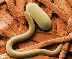
Nematodes, more commonly known as roundworms or threadworms, are found parasitizing plants, humans, and other animals and can also inhabit soil, freshwater, and saltwater habitats. Ranging from 0.3mm to 8.5m in length, the largest roundworm recorded is Placentonema gigantisma from the placenta of a sperm whale.
Nematodes are unsegmented, bilaterally symmetrical, pseudocoelomate animals, and the mouth can contain teeth or stylets used to penetrate the host.
Although they lack a circulatory and respiratory system, they have a digestive system in which the food is moved through the tract via internal/external pressures and body movement (as opposed to muscles).
Most nematodes are not parasitic, but those that are usually have complicated life cycles involving several hosts or locations within a host.
Anguillicola crassus: Classification and Morphology
1. Classification:
Kingdom: Animalia
Phylum: Nematoda
Class: Chromadorea
Order: Spirurida
Family: Anguillicolidae
Genus: Anguillicola
Species: A. crassus
2. Size Range: Male parasites range from 20-60µm in length, with females measuring 47-72µm. Body width ranges from 0.9-2.8µm for males and 3-5.6µm for females.
3. Life Stages: Eggs are released from the digestive system of the definitive host. Larvae attach to the substratum by their hooked tails. Transmission out: Larvae are ingested by the intermediate host. Free-living larvae are ingested by the intermediate host.
Often a copepod or other crustacean, particularly Cyclops vicinus and C. albidus. The intermediate host is eaten by the definitive host. Anguilla sp. (Anguilla anguilla in Britain).
Infected eels develop a disease called ‘Anguillicolosis,’ which causes hemorrhagic lesions, fibrosis, collapsed swim bladders, and inflammatory reactions.
The adult nematode has a soft, wrinkled outer cuticle with a small circular mouth opening, which is surrounded by 4 dorsolateral and ventrolateral papillae and 2 small lateral amphids.
Acanthocephala: Morphology and Taxonomy in Fish Health
Acanthocephalans, otherwise known as spiny- or thorny-headed worms, are highly specialized intestinal parasites that use a spiny proboscis to penetrate and attach to host tissues. Nutrient uptake occurs directly through the body surface of the parasite, as it lacks both a mouth and an alimentary canal.
Acanthocephalan life cycles are usually complex and may involve a number of hosts, including invertebrates, fishes, amphibians, birds, and mammals. This phylum is currently divided into three classes: Archiacanthocephala (with terrestrial life cycles), Palaeacanthocephala (aquatic life cycles with fish, seals, or water birds being the final hosts), and Eoacanthocephala (aquatic life cycles with fish, reptiles, and amphibians being the final hosts).
Species of Acanthocephala Found in Freshwater Fish
Species found in freshwater fish in Britain and Ireland include: Acanthocephalus clavula, A. lucii, A. anguillae, Echinorhynchus borealis, E. clavula, E. salmonis, E. truttae, Neoechinorhynchus rutili, and Pomphorhynchus laevis.
Pomphorhynchus laevis: Classification and Morphology
1. Classification:
Kingdom: Animalia
Phylum: Acanthocephala
Class: Palaeacanthocephala
Order: Echinorhynchida
Family: Pomphorhynchidae
Genus: Pomphorynchus
Species: P. laevis
2. Size Range: Size varies between 4 and 30mm and generally increases with age. Acanthocephalans are dioecious: males are usually smaller at 6-16mm, with females being 10-30mm. Eggs are 110-121 x 10^-19µm.
Read Also: 13 Medicinal Health Benefits of Chimaphila umbellata (Pipsissewa)
Trematodes: Morphology and Taxonomy in Fish Health
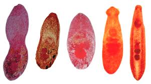
Trematodes, members of Phylum Platyhelminthes, also called flukes, cause a variety of clinical infections in humans worldwide. The parasites are named trematodes because of their conspicuous suckers, which are the organs of attachment (trematode means “pierced with holes”). All of the flukes that cause infections in humans are contained in the group called “digenetic trematodes.”
Depending on their habitat in the infected host (generally a vertebrate), flukes can be classified as blood flukes, liver flukes, lung flukes, and intestinal flukes. Flukes causing most human infections are Schistosoma species (blood fluke), Paragonimus westermani (lung fluke), and Clonorchis sinensis (liver fluke).
Some less clinically important flukes are Fasciola hepatica and Opisthorchis viverrini, which are liver flukes, and Fasciolopsis buski, Heterophyes heterophyes, and Metagonimus yokogawai, which are all intestinal flukes.
Features of Trematodes
The sexes of the parasites are not separate (monoecious). In other words, they are mostly hermaphroditic, with the male and female reproductive organs existing complete in each fluke. One exception is the schistosomes, which are dioecious.
The flukes are oviparous and lay diagnostically operculated eggs. Once again, an exception is schistosome eggs, which are not operculated. They are unsegmented, dorso-ventrally flattened, and leaf-shaped.
The alimentary canal is incomplete, with the anus being absent. The excretory system is bilaterally symmetric. They bear two suckers, one on the ventral surface of the body (ventral sucker) and one around the mouth (oral sucker). These serve as organs of attachment for the fluke.
Taxonomy of Trematodes
Class: Trematoda
Subclass: Digenea (the digenetic trematodes)
Order: Opisthorchiformes
Family: Opisthorchiidae
Species: Clonorchis sinensis
Leeches: Morphology and Taxonomy in Fish Health
Leeches have so far only been reported from a few fish in Africa, including Bagrus docmac, Barbus altianalis, B. tropidolepis, carp, and Protopterus aethiopicus.
However, leeches apparently attack a wider range of fish (Claridae, Synodontidae, Mormyridae, and Cichlidae) and in a greater number of water systems, as is evident from the distribution of leech-transmitted trypanosomes in African fish.
Most records of leeches removed from fish in Africa are of Batrachobdelloides tricarinata. This leech occurs from the Jordan system in Israel, infecting Clarias lazera, throughout tropical West and East Africa to Zululand in Southern Africa (Oothuizen, 1989).
Piscicolid leeches are common parasites of Mugilidae in the riverine-estuarine system of the southern Cape Province in South Africa.
Description and Taxonomy of Leeches
Leeches feeding on fish are Rhynchobdellae and belong either to the Glossiphoniidae or the Piscicolidae (Mann, 1962). Most named records of Glossiphoniid leeches from African fish and many of those found free in the habitat were proven to be synonymous with B. tricannata.
There is one record of another fish-feeding glossiphonid, Hemiclepsis quadrata (Moore, 1939), from Ethiopia (Oosthuizen, 1987). Apart from the piscicolid leeches (as yet undescribed) of Cape grey mullets, the only other African record of a piscicolid leech is of a species of Phyllobdella removed from Barbus (Moore, 1939).
Rhynchobdellae have a small pore-like mouth on the oral sucker from which a proboscis may be protruded; no jaw is present, and the blood is transparent (Gnathobdellae, which feed on higher vertebrates, have a large mouth with jaws and red blood). Differentiation even between Piscicolidae and Glossiphoniidae is not easy for the non-expert:
1. Glossiphoniidae: The body at rest is depressed, not divided into distinct anterior and posterior regions; the head is usually much narrower than the body, with an anterior sucker either indistinguishable or only slightly distinct from the body. There are usually 3 annuli per segment in the mid-body region, and eyes are confined to the head.
2. Piscicolidae: The body at rest is cylindrical and (especially when contracted) usually divided at segment XIII into distinct anterior and posterior regions. The head sucker is usually distinctly marked off from the body, which usually has more than three annuli per segment. Simple eyes may be present on the head, neck, and posterior sucker.
Experts would prefer leeches to be live and fixed to their own specifications. Leeches can survive for a considerable time, even when mailed in a vial inside wet cotton wool. If fixed, it is best in 70% ethanol, and preferably, the leech should be relaxed first with menthol, ether, or by refrigeration, sometimes, if not too small, under glass slide pressure.
Lymphocystis Virus: Morphology and Taxonomy in Fish Health
1. Species Affected: A variety of marine and freshwater fish; in Africa, known only from cichlids, including species of Tilapia, Oreochromis, and Haplochromis.
2. Geographic Range: In cichlid fish in Lakes Victoria (Nyanza) (Oreochromis variabilis and Haplochromis spp.), in Lake George (H. elegans), and L. Kitangiri (Tilapia amphimelas and O. esculentus) in East Africa.
Description, Taxonomy, and Diagnosis
Infection is manifested in one to numerous dermal clusters of rounded pustules or wart-like growths. Histological sections reveal aggregates of grossly hypertrophic cells (in cichlids, 200–330 µm in diameter), enclosed within a thick hyaline (eosinophilic) wall and an extremely large nucleus and nucleolus.
Cytoplasm contains basophilic (DNA) inclusions (numerous, small-rounded in cichlids) and vacuoles. Lymphocystis viruses are large (160–300 nm), icosahedral, DNA viruses (iridovirus-like) replicating within the cytoplasmic inclusions and are released into the cytoplasm to form regular arrays.
Good taxonomy is essential for ecological, biogeographical, and evolutionary studies of any group of organisms. Fish serve as hosts to a range of parasites that are taxonomically diverse and that exhibit a wide variety of life cycle strategies.
Whereas many of these parasites are passed directly between ultimate hosts, others need to navigate through a series of intermediate hosts before reaching a host in (or on) which they can attain sexual maturity.
Do you have any questions, suggestions, or contributions? If so, please feel free to use the comment box below to share your thoughts. We also encourage you to kindly share this information with others who might benefit from it. Since we can’t reach everyone at once, we truly appreciate your help in spreading the word. Thank you so much for your support and for sharing!
Read Also: How To Educate Yourself On Climate Change

