In this article, reproduction in domestic livestock will be studied. In fact, the article presents both the macroscopic and microscopic features of the reproductive tract of a typical farm animal such as a bull.
Definition of Reproduction in Farm Animals
Reproduction is defined as the process, sexual or asexual, by which animals and plants produce new individuals. Reproduction in animals normally involves heterosexual mating, conception, pregnancy, parturition and lactation.
In this process, conception occurs as a result of the fusion of the male and female gametes, namely spermatozoon and ovum respectively, in a process known as fertilization. For animals to reproduce, they must first attain puberty or reproductive age, from when they become capable of gamete production.
Reproduction in animals involves close coordination or synchronization of various physiological events, and this is largely achieved through the actions of reproductive hormones.
Read Also: Feed Lot Fattening of Rams Practice
Male Reproductive Anatomy (Gross Structures)

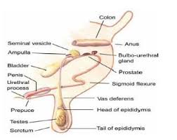
These structures are illustrated in Fig. 1 below.
1. Scrotum (Protective Sac for Testes):
A pendulous sac or pouch containing the testes. It consists of:
i. Skin: outer covering for protection
ii. Dartos muscle: sheet of muscle under the skin of the ventral half of the scrotum
Function: Contracts in cold weather to pull the testes closer for warmth and relaxes in hot weather to allow cooling.
2. Testes (Sperm‑ and Hormone‑Producing Organs):
Two in number, located side by side; oval or rounded shape (125 × 75 × 75 mm), suspended by the spermatic cord.
Function: Produce sperm (male gamete) and secrete the hormone testosterone.
3. Epididymis (Sperm Transport and Maturation Duct):
Elongated convoluted duct comprising three sections:
i. Head (Caput): flattened portion attached to the anterior third and dorsal surface of each testis
ii. Body (Corpus): cord‑like portion loosely attached along the posterior border of the testis
iii. Tail (Cauda): bulging terminal portion on the ventral surface of the testis
Function: Transport, maturation, concentration and storage of sperm.
4. Vas Deferens (Sperm Conduit):
Single tube originating at the tail of the epididymis, passing through the inguinal ring into the body cavity, over the dorsal surface of the bladder into the anterior urethra. Duct measures approximately 2 mm × 50 cm, with an enlarged ampulla before entering the urethra.
Function: Transport and some storage of sperm, plus active muscular contractions during coitus.
5. Spermatic Cord and Associated Muscles:
Includes the inguinal ring, cremaster muscle and connective tissues that attach and support the external genitalia.
Function: Support the testes and regulate their position for optimal temperature.
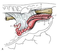
6. Accessory Glands (Seminal Vesicles, Prostate, Bulbourethral):
i. Vesicular glands: paired lobulated glands dorsal and anterior to the bladder‑urethra junction; secrete fluid rich in fructose.
ii. Prostate: two‑lobed body across the dorsal urethra; secretes fluid to clean and lubricate the urethra.
iii. Bulbourethral glands: embedded paired glands at the root of the penis; secrete fluid to cleanse and lubricate the urethra pre‑ejaculation.
Male Copulatory Organ

1. Penis (Organ of Copulation and Sperm Deposition):
Comprises root (bulbourethral muscle area), body or shaft, sigmoid flexure (S‑curve for length adjustment), and glans (soft terminal portion).
Function: Transport, insertion and deposition of sperm into the female reproductive tract.
2. Retractor Penis Muscles:
Paired muscles attached to the sigmoid flexure and sacral area.
Function: Retract the penis when relaxed and permit extension during copulation.
3. Prepuce and Sheath:
Foreskin sheath with many folds and coarse hairs; secretion and debris form smegma.
Function: Protect the glans and maintain hygiene.
Male Reproductive Anatomy (Microscopic Features)
1. Tunica Vaginalis Propria and Albuginea (Testis Coverings):
i. Vaginalis propria: outer connective tissue, continuation of peritoneum.
ii. Albuginea: white connective tissue layer beneath the tunica vaginalis propria; provides support.
2.Seminiferous Tubules (Sites of Spermatogenesis):
Coiled tubules composed of basement membrane and sustentacular (Sertoli) cells that nourish developing spermatogenic cells.
Function: Support and nurture spermatids as they develop into spermatozoa.
3. Spermatogenic Cells and Stages:
i. Spermatogonia: primitive cells on basement membrane; give rise to spermatocytes.
ii. Primary and secondary spermatocytes: stages leading to haploid spermatids.
iii. Spermatids: form via meiotic division and transform into spermatozoa through spermiogenesis.
4. Spermatozoon (Mature Germ Cell):
Motile cell with distinct head, body and tail; released into the lumen after maturation.
5. Interstitial (Leydig) Cells:
Large cells between seminiferous tubules; produce testosterone in response to pituitary signals.
6. Supporting Structures:
Rete testes, efferent ducts, serosa, epithelium, blood vessels, lymphatics and nerves all facilitate sperm transport and testicular function.
Read Also: General Principles of Goat Production
Female Reproductive Anatomy (Gross Structures of the Cow)

1. Ovary (Egg‑Producing Organ):
Two oval structures (30 × 20 × 40 mm) supported by broad ligaments.
Function: Produce ova and secrete female sex hormones (estrogens and progestogens).
2. Oviduct (Site of Fertilization and Transport Tube):
Two tortuous tubes (~100 mm) suspended by broad ligaments.
Function: Convey sperm and ova, site of fertilization, and secrete nurturing fluids.
3. Uterus (Incubation and Nutrient Supply Organ):
Y‑shaped with two horns and one body (37.5 cm long); supported by broad ligament.
Function: Aid sperm transport, secrete uterine fluids, incubate the embryo and supply nutrients.
4. Cervix (Protective Neck of the Uterus):
Single thick‑walled tube (100 mm × 25 mm).
Function: Permit sperm passage, secrete mucus, seal the uterus in pregnancy, and allow fetal passage at birth.
5. Vagina (Birth Canal and Copulatory Receptacle):
Tubular structure (200 mm × 50 mm) extending from cervix to external opening.
Function: Receive the penis, secrete mucus and serve as passage for the fetus.
6. Vulva (External Urogenital Opening):
Labial structures forming the external genital lips.
Function: Passageway for urine, receptor for the penis during copulation, and exit for the fetus at birth.
Female Reproductive Anatomy: Microscopic Structures of the Cow

As can be seen from Fig. 4, the microscopic structures of the cow’s ovary and tubular tract exhibit numerous specialized features.
1. Peritoneum: thin membrane of connective tissue covering the ovary
Helps maintain shape and structure.
2. Germinal epithelium: single layer of cuboidal cells beneath the peritoneum
Is sometimes difficult to distinguish ; gives rise to the female germ cell, the ovum.
3. Tunica albuginea: connective tissue membrane beneath the germinal epithelium
Maintains ovarian structure.
4. Cell nest: invagination of a cord of germinal epithelial cells
One cell will differentiate into an ovum ; the others become supportive cells.
5. Stroma: mass of connective tissue, blood vessels, nerves and other inclusions
Provides a structural base to the organ.
6. Primary follicle: ovum surrounded by a single layer of follicular cells
Usually found near the ovarian periphery.
7. Secondary follicle: ovum surrounded by two layers of follicular cells
Located deeper in the stroma.
8. Growing follicle: follicular cells separate forming an antrum filled with fluid
The ovum moves to the periphery inside the follicle ; theca externa and theca interna organize outside the granulosa cells.
9. Mature (Graafian) follicle: antrum enlarges, protruding on the surface as a blister
From outside inward observe:
i. Theca externa: connective tissue support
ii. Theca interna: dense connective tissue and estrogen source, highly vascular
iii. Basement membrane: thin connective tissue layer
iv. Granulosa membrane: cuboidal cells lining the antrum and source of estrogen
v. Antrum: cavity filled with estrogen‑rich follicular fluid
vi. Cumulus oophorus: granulosa cell mound supporting the ovum within the antrum
vii. Corona radiata: spindle‑shaped cells adjacent to the ovum
viii. Zona pellucida: transparent, non‑cellular membrane beneath the corona radiata
ix. Perivitelline space: gap between the zona pellucida and vitelline membrane housing the polar body
x. Vitelline membrane: cell membrane of the ovum cytoplasm
xi Vitellus: cytoplasm containing nutrients and inclusions
xii. Nucleus: chromosome‑bearing portion of the ovum
xiii. Polar body: extruded nucleus with half the chromosomes, located in the perivitelline space
xiv. Hillock: stalk of granulosa cells supporting the ovum cell mass
10. Corpus hemorrhagicum: ruptured follicle filled with blood
Granulosa cells begin differentiating into larger, rounded lutein cells ; this transient stage is rarely seen in sections.
11. Corpus luteum: mass of rounded luteal cells of varying sizes
Theca layers are less distinct ; luteal cells produce progesterone.
12. Corpus albicans: connective tissue and degenerating luteal cells
Old corpus luteum resorbed into the stroma, appearing as a small, dense connective tissue mass.
13. Atretic follicle: degenerating follicle invaded by connective tissue
Numerous atretic follicles exist in varying stages of resorption; the ovum may sometimes remain entrapped.
14. Cystic follicle: intact follicle that has not ruptured
Low LH or mechanical causes may be responsible ; theca and granulosa continue estrogen secretion, causing continuous or intermittent estrus.
15. Persistent corpus luteum: corpus luteum that does not regress
Gross and microscopic structure resembles a mature corpus luteum ; associated with progesterone–FSH imbalance, leading to pseudopregnancy or anestrus.
Female Reproductive Anatomy: Microscopic Structures of the Tube and Uterus
1. Oviduct
i. Serosa: outer connective tissue cover — shiny and tough, giving shape and strength.
ii. Muscularis: smooth muscle layer beneath the serosa — longitudinal fibers outer, circular/spiral inner; peristaltic contractions move sperm and ovum toward the tubo‑uterine junction.
iii. Mucosa: lining of many longitudinal folds — more abundant near the ovary, decreasing toward the uterus, covered by ciliated columnar, secretory (goblet) and nonsecretory cells.
2. Uterus
i. Serosa: outer connective tissue cover — shiny and tough, gives shape and strength.
ii. Myometrium: smooth muscle layer — outer longitudinal and inner circular/spiral fibers contract under estrogen to move sperm.
iii. Endometrium: mucosal layer
Stroma: structural connective tissue
iv. Glands: tortuous spiraled glands in the stroma — secrete fluids for sperm/ovum transport and early embryo nutrition.
v. Epithelium: columnar cells on a basement membrane — varies from simple to pseudostratified, sloughs during pregnancy in cow and ewe.
3. Cervix
i. Serosa: outer connective tissue cover — shiny and tough.
ii. Muscularis: dense muscle and connective tissue — opens under estrogen for sperm and fetus passage, closes under progesterone to seal the uterus.
iii. Mucosa: many folds giving a fern‑like cross‑section — crypts lined with secretory cells, outer folds covered with columnar epithelium; secretion thin under estrogen, tenacious under progesterone.
4. Vagina
i. Serosa: outer connective tissue cover — shiny and tough.
ii. Muscularis: inner circular and outer longitudinal — less developed than uterus.
iii. Mucosa: stratified squamous epithelium — thickness and activity hormone‑dependent; deep papillae protect against abrasion.
5. Vulva
i. Stroma: connective tissue, fat cells and vestibular glands — supports the structure.
ii. Mucosa: thick stratified squamous epithelium with deep papillae — protects the vestibule and supports lubrication.
Reproduction and Related Disciplines in Agriculture
A proper understanding of animal reproduction involves knowledge of reproductive physiology, endocrinology, environmental physiology, cell biology, immunology, genetics, biochemistry, sociology, reproductive diseases, embryology, obstetrics and related fields.
In agriculture, animal production centers on reproduction: eggs and milk derive directly from reproductive processes, and meat production depends on offspring born, raised and fattened for slaughter.
This article provides a concise description of the gross and microscopic reproductive tracts of male and female farm animals, using cattle as a model, and highlights the importance of interdisciplinary understanding in animal reproduction.
Do you have any questions, suggestions, or contributions? If so, please feel free to use the comment box below to share your thoughts. We also encourage you to kindly share this information with others who might benefit from it. Since we can’t reach everyone at once, we truly appreciate your help in spreading the word. Thank you so much for your support and for sharing!

