Epidemics of bacterial diseases are common in dense populations of cultured food or aquarium fish. Predisposition to such fish diseases outbreaks frequently is associated with poor water quality, organic loading of the aquatic environment, handling and transport of fish, marked temperature changes, hypoxia, and related stressful conditions.
High concentrations of waterborne bacteria are normally found in ponds and aquaria. Many of these bacteria are opportunistic facultative pathogens, which are activated by an adverse environment, a debilitated host, or a primary pathogen.
In sharp contrast, obligatory bacterial pathogens of finfish require the presence of fish for replication and are unable to survive alone for long in the aquatic environment.
Most bacterial pathogens of fish are aerobic gram-negative rods. Diagnosis is by isolating the organism in pure culture from infected tissues and identifying the bacterial agent.

Stained Gram-positive bacteria viewed at high magnification

Stained Gram-negative bacteria viewed at high
Eye Disease
Fish are prone to developing bacterial infections in their eyes. Several different bacteria can grow and have devastating effects on a fish, as well as spread to the other fish in your aquarium.
Luckily, many infections can easily be cured by adding medicated drops to your fish pond or tank.
Eye diseases and infections in fish can be caused by a variety of factors, including fungus, bacteria, parasites, a kidney infection or a protozoan infestation.
Fish can exhibit a variety of symptoms when suffering from an infection or bacterial disease.
These include a cottony growth around the eyes, cloudiness in the eyes, eyes that appear to be popping out of socket and film on the eyes. Diagnosing the cause of the fish’s eye ailment will depend on the symptoms.
Cottony white growth around the eye is caused by a fungus and a cloudy iris is caused by bacteria. If the entire eye looks cloudy, it is a protozoan infestation, and if the eye appears to be popping out, it is kidney failure.
A liquefactions infects the eyes of silver carp. The cornea of the eyes gets vascularized leading to opacity and complete necrosis and even mass mortality of fish has been recorded.
Investigations have isolated staphylococcus aureus from the affected eyes of diseases fish.
Treatment
A fungal infection is treated using sulfate drops, which you can administer to the tank water as directed. Other infections are treated with antibiotic drops, which also are administered to the water.
The affected fish can be treated with other fish in the tank; the antibiotic drops will prevent the infection from spreading to them. Chloromycetin bath @ 8 – 10mg/L has been found effective in controlling the disease at an early stage.
Disinfecting the environment with Potassium permanganate at a dose of 0.1 ppm followed by liming at 300 ppm check the disease.
Ulcerative disease bilateral ulcerations of the opercula and the head in catfish are observed in ulcerative disease. In most cases, A. hydrophilic could be isolated, although several other bacterial forms were also present as secondary invaders.
Enteric Septicemia of Catfish (ESC)
ESC caused by the gram negative bacterium Edwardsiella ictaluri, is one of the most important diseases of farm-raised channel cat- fish (Ictalurus punctatus).
ESC accounts for approximately 30 percent of all disease cases sub- mitted to fish diagnostic laboratories in the southeastern United States. In Mississippi, where channel catfish make up the majority of case submissions, it has been reported at frequencies as high as 47 percent of the yearly total.
Economic losses to the catfish industry are in the millions of dollars yearly and continue to increase steadily with the growth of the industry. The disease was similar to another disease of catfish caused by the gram negative bacterium Edwardsiella tarda, but differed in several characteristics.
ESC was described in a published account in 1979 and the causative bacterium was described as a new species in 198.1
Clinical Signs and Diagnosis
Behavior Catfish affected with ESC often are seen swimming in tight circles, chasing their tails. This head-chasing-tail, whirling behavior is due to the presence of the Edwardsiella ictaluri in the brain.
Affected fish also sometimes hang in the water column with the head up and tail down. In addition, catfish with ESC tend to stop eating shortly after becoming infected.
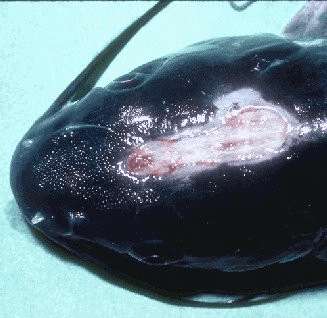
External Signs (ESC) affected catfish frequently have red and white ulcers (ranging from pinhead size to about half the size of a dime) covering their skin (Fig. 1); pinpoint red spots (called petechial haemorrhages) especially under their heads and in the ventral or belly region (Fig. 2); and longitudinal, raised red pimples at the cranial foramen between the eyes (Fig. 3) that can progress into the hole-in- head condition.
Internal build-up of fluid can lead to a swollen abdomen and exophthalmia (popeye) (Fig. 4).
Internal Signs
Clear, straw-color or bloody fluid is often present in the fish’s body cavity. The liver typically has characteristic pale areas of tissue destruction (necrosis) or a general mottled red and white appearance (Fig. 5).
Petechial hemorrhages can be found in the muscles, intestine and fat of the fish. The intestine is also often filled with a bloody fluid.
Treatment
Treatment of ESC can be approached in a variety of ways. A good pond manager makes daily observations on feeding response, behavior and mortality, thus making an early diagnosis possible.
Traditionally catfish infected with ESC are treated with feeds containing antibiotics. First, samples of sick fish should be submitted to a fish diagnostic laboratory for a complete diagnosis.
The causative bacterium can then be isolated and tested for antibiotic sensitivity. Fish should be treated as soon as a diagnosis has been made because fish progressively reduce feed intake during an infection, making medicated feed treatments less effective.
Columnaris Disease
Flexibacter Columnaris (Columnaris disease or Saddle back disease) Columnaris, first described by Herbert Spencer Davis in 1922, is one of the oldest known diseases of warm water fish. References to the disease can be confusing.
The causative bacterium has been referred to by different names including Bacillus columnaris, Flexibacter columnaris, Cytophaga columnaris, and most recently Flavobacterium columnare.
Columnaris disease is the second leading cause of mortality in pond raised catfish in the southeastern United States. It is second only to enteric septicemia of catfish (ESC) caused by the bacterium Edwardsiella ictaluri.
Most species of fish are susceptible to columnaris following some type of environmental stress and when water temperatures are in the upper part of their preferred temperature range.
Read Also : Bacterial Fish Diseases and Control Measures
The disease commonly occurs in channel catfish when water temperatures are in the range of 25 to 32oC (77 to 90o F) in the spring, summer and fall.
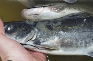
Clinical signs or symptoms Fish with columnaris usually have brown to yellowish-brown lesions (sores) on their gills, skin and/or fins.
The bacteria attach to the gill surface, grow in spreading patches, and eventually cover individual gill filaments (Fig. 1).
This results in cell death. Portions of the gills are eroded by protein and cartilage-degrading enzymes produced by the bacteria. Skin lesions produced by columnaris initially are very shallow and may appear as an area that has lost its natural shiny appearance.
More advanced lesions may be round or oval in shape, yellowish-brown in color, with an open ulcer in the center. A characteristic lesion produced by columnaris is a pale white band encircling the body, often referred to as saddleback condition (Fig. 2).
As the infection progresses, a yellowish-brown ulcer often is found in the center of the “saddle.” Additionally, it is not unusual to find a yellowish-brown, mucus-like growth of columnaris bacteria inside the fish’s mouth (Fig. 3).
The brownish coloration is usually due to mud and detritus particles trapped in the slime produced by the bacteria. When grown in the laboratory the bacteria produce a yellow pigment.
Gram negative slender rods (3-8 microns). The disease is a serious disease of young salmonids, catfish and many other fish. This is a highly communicable disease. Cause external lesions over the body surface.
The causative organism has been identified as Flexibactercolumnaris . Lesions usually first appear as small white spots on the caudal fin and progresses towards the head.
The caudal fin and anal fins may become severely eroded. As the disease progresses, the skin is often involved with numerous gray white ulcers. Gills are a common site of damage and may be the only affected area.
The gill lesions are characterized by necrosis of the distal end of the gill filament that progresses basally to involve the entire filament.
Flexibactercolumnarisinfections are frequently associated with stress conditions. Predisposing factors for Columnaris disease are high water temperature (25°C-32°C.), crowding, injury, and poor water quality (low oxygen and increased concentrations of free ammonia).
Flexibacter maritimus: cause similar problems in salt- water environment. Flexibacterpsychrophilus causes Cold Water Disease or Peduncle disease. Fish develop dark skin, hemorrhage at the base of fins, and anemia with pale gills with increase mucus.
Hemorrhage into the muscles is common. Periostitis of cranial and vertebral bones is common in chronic cases. A chronic meningoencephalitis occasionally is observed with abnormal and erratic swimming.
Treatment for external columnaris infection includes treating the culture water with therapeutic chemicals legal for use on food fish. Potassium permanganate (KMnO4) is a commonly used therapy.
| 100 lbs | 5.00 g/lb. | 100 lbs 0.5 – 0.75 % |
| 50 lbs. | 2.50 g/lb. | 1.0 – 1.5 %. |
| 25 lbs. | 1.25 g/lb | 2.0 – 3.0 % |
Bacterial Gill Disease
Bacterial gill disease is caused by a variety of bacteria. Flexibacter columnaris, Flexibacter psychrophilus, Cytophaga psychrophila and various species of Flavobacterium (all are gram negative rods) are the primary bacteria involved in this disease.
Fry are the most susceptible to the disease, however, all ages may be affected. Clinically the fish become anorectic, and face the water current. Prominent hyperplasia (mucus and epithelial) of the gills is evident on gross and microscopic examination.
Microscopically one observes proliferation of the epithelium that result in clubbing and fusion of the lamella. Necrosis of the gill lamella occurs in serious cases.
Overcrowding, accumulation of metabolite waste products (particularly ammonia), organic matter in the water, and an increase in water temperature may all be predisposing factors.
Topical application of potassium permanganate or short bath in 500ppm of Potassium permanganate has been found to be very effective in completely curing the disease.
Proliferative Gill Disease (PGD)
Proliferative gill disease (PGD) has become common in farm raised channel catfish. It can kill a few dozen fish over several days, or up to 100 percent of the fish in less than 3 days. Recurrence in the same pond is rare.
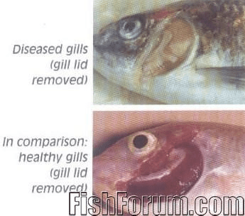
This disease causes catfish to suffocate because of the severe damage to the gills. Swelling and a red and white mottling of the gills gives them a raw hamburger appearance, and many refer to PGD as hamburger gill disease.
Proliferative Gill Disease(PGD)
Clinical Signs and Diagnosis
Proliferative gill disease occurs most often in the spring, but it can occur in the fall at water temperatures between 59 and 72o F (15 to 22oC). It sometimes occurs in winter; PGD mortalities have been reported at 43o F (6o C). Though the disease seldom occurs in the summer, deaths have been reported at 92oF (33o C).
Even before the disease occurs, signs of PGD can be seen in gills viewed under the microscope. As with other diseases, a common early sign of a PGD outbreak is a reduction of feeding activity by the fish.
As the disease progresses, the catfish congregate in the water flow behind an aerator or at incoming water. Fish may also swim listlessly at the water’s surface and then lie in shallow water along the edge of the pond before they die.
They may die even when dissolved oxygen concentrations are at levels high enough for healthy fish, because the affected gills cannot remove sufficient oxygen from the water.
The skin of catfish affected with PGD appears healthy, and while PGD occasionally is found in internal organs (liver, kidney, spleen and brain), it primarily affects the gills.
The gills swell and become mottled red and white in appearance, similar to raw hamburger meat. In advanced stages, the gill filaments do not lie flat and filaments on one gill arch are not distinct from filaments on other arches. The gills often look mashed and may bleed when touched or when the fish are simply lifted from the water.
Microscopic examination at 40X magnification reveals extreme swelling of the gills caused by an abnormally large number of cells at the outer edge of the gill filaments. These swollen areas often appear white.
PGD causes swelling and red and white mottling of catfish gills, giving them a raw hamburger appearance. Some parts of the gill filaments look red because blood cells are pooled in ruptured or dilated capillaries.
The gill filaments may become shorter and wider with rounded or squared tips. The cartilage supporting the gill filament appears as a dark gray band along the side of each filament and may have notches, breaks and gaps.
These characteristics are also much more obvious when examined under 40X magnification than at higher magnifications, and are the best features for making an early, presumptive diagnosis. The lesions in the cartilage can be occupied by the parasite that causes PGD.
Breaks and gaps in the filaments’ supporting cartilage cause the gills to lose their well-defined structure and collapse onto each other, giving the mashed appearance. Parasite cysts are only occasionally seen in wet mounts under the microscope and appear as small, indistinct, round units.
PGD diagnosis is confirmed by histology procedures where the parasite can be seen as a blue stained “cluster of grapes” in very thinly cut sections of gill tissue. Cause and disease course Most scientists believe that a sporozoan, probably the myxosporean parasite Aurantiactinomyxon sp., is the causative agent of PGD.
Evidence suggests that an oligochaete worm (Dero digitata) that lives in the mud and grows up to 1/2 inch in length is the invertebrate host.
The PGD organism is thought to develop in the worm, which releases infective spores capable of penetrating and infecting the gills of channel catfish.
Most parasites inflict less damage to their natural hosts, and mature spores are usually found in the host tissue. Most of the gill damage is thought to be caused by an inflammatory response of the fish to the parasite.
Following is a possible life cycle of PGD:
Mature parasites (possibly Aurantiactinomyxon sp.) or an infective stage are released from the fish host.
The invertebrate host (probably Dero digitata) becomes infected.
The parasite develops in the invertebrate host.
An infective stage of the parasite is released from the worm.
This infective stage penetrates and infects the catfish gill tissue.
There has been an ongoing controversy among researchers and diagnostic workers about the occurrence of this disease in new ponds built or reworked within the last 3 years as compared with older ponds.
Although PGD occurs in older ponds, it seems to appear more often in new ponds, perhaps because they support larger populations of Dero worms.
Treatment and Prevention
Though no treatments or preventive methods for PGD have been scientifically validated, there are some treatments that appear effective. Since fish infected with PGD suffer gill damage and are less able to obtain oxygen from water, aeration should be used when dissolved oxygen concentrations are low or marginally low. Another option is to quickly harvest and process PGD infected fish.
However, many fish may die during harvest and have to be discarded; those making it to the processing plant alive can be quickly processed and pose no danger to the human consumer.
Edwardsiellosis
It is a septicaemic disease affecting brood fish population. Edwardseilla tarda has been isolated from the diseases fish showing anaemia, cutaenous lesions and gas filled abscesses in the muscle.
Edwardsiella tarda (Edwardsiella septicemia). Gram negative motile pleomorphic curved rod. The disease affects primarily channel catfish but also observed in goldfish, golden shiners, largemouth bass, and the brown bullhead.
This organism is the most serious disease involving the eel culture of Asia. The lesions are similar to A. hydrophila with small cutaneous ulcers and hemorrhage observed both in the skin and muscle.
Muscle lesions often develop into large gas filled (malodorous) cavities. Diseased fish lose control over the posterior half of their body yet continue to feed.
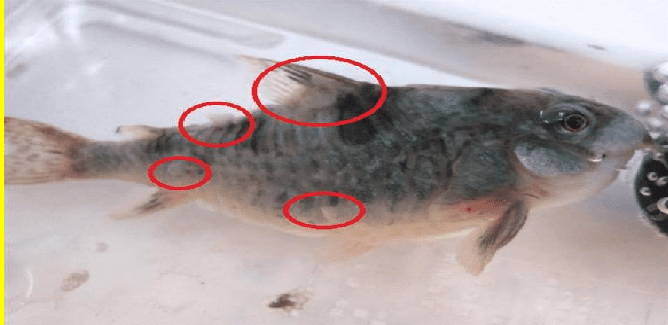
Edwardsiella ictaluri (Enteric septicemia of catfish): Gram negative motile pleomorphic curved rod. Disease affects primarily fingerlings and yearling catfish. Clinical signs of enteric septicaemia of catfish closely resemble those of other systemic bacterial infections. The most characteristic external lesion is the presence of a raised or open ulcer on the frontal bone of the skull between the eyes (Hole in the head disease).
Although treatment with idophor has been found to be effective, water quality improvement in the hatchery is the most essential component for keeping the disease away.
Enteric Septicemia of Catfish
Epizootic ulcerative syndrome (EUS)
Epizootic ulcerative syndrome (EUS) has become a matter of great concern not only among fisherman and fish farmers, but also among general public, entrepreneurs, administrators and planners.
One common feature of the disease is that it initially affects the bottom dwelling species like murrels followed by catfishes, weed fish es and IMC.
The lesions start as small grains to pea sized hemorrhagic spots over the body which ultimately turns into big ulcers of the size of a coin with greyish slimy central necrotic areas surrounded by a zone of hyperemia.
The disease affects to such an extent that they starts rotting while still alive and eventually die. A number of bacteria viz.A. hudrophila, A. punctata, Flavobacterium sp.,Pseudomonas sp., Edwardsiella tarda, Vibrio parahaemlyticus and Streptococcos sp. have been isolated from the affected specimens.
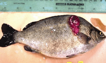
Vibrio
Histopathological studies revealed complete loss of epidermis in the ulcerative area of the skin where the dermis and hypodermis showed characteristic granulomatous changes. Besides bacteria, virus, fungus and parasites were also reported to be associated with EUS.
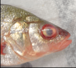
The eyes and fins caused by a bacterium called vibrio
Many antibiotics, sulfonamides, chemicals herbal preparations etc. have been advocated as preventive and curative measures.
Lime was accepted widely among fish farmers of the country until the formulation of CIFAX, therapeutics developed by Central Institute of Freshwater Aquaculture (CIFA). Marked improvement of the ulcerative condition is noticed within seven days of application of the medicine and the ulcers are healed up within 10 -14 days.
Streptococcus iniae
Beta-hemolytic Streptococcus (Note: Beta hemolysin may not be present in culture media in all cases leading to the possible believe that this bacteria is a non-pathogen.). Disease of tilapia, hybrid striped bass and rainbow trout. Major problem in the tilapia industry.
Streptococcus iniae presents either as an acute fulminating septicemia or in a chronic form limited primarily to the central nervous system. The septicemic form may present with hemorrhage of the fins, skin, and serosal surfaces.
Ulcers may appear. Microscopically, one observes a meningoencephalitis, polyserositis, epicarditis, myocarditis and/or cellulitis. Cocci/diplococci are present in the inflammation. In the chronic form, granulomas or granulomatous inflammation are evident in the liver, kidney, and brain (meningoencephalitis).
In the chronic disease, the brain is the best organ to culture. Streptococcus iniae is a problem primarily of closed recirculating culture system. Probably associated with overcrowding and poor water quality – high nitrates.
Depopulation, disinfection and restocking with disease free fish are the best means of elimination of the organism. The bacteria is known to be a zoonotic agent. Individuals who have handled infected fish have developed cellulitis of the hands and endocarditis.
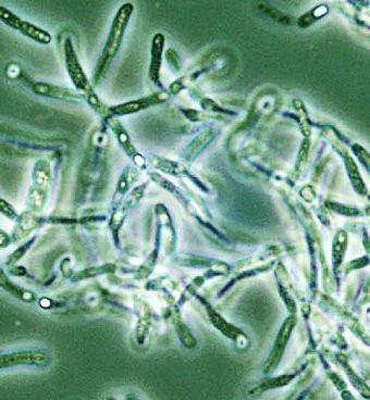
Streptococcus iniae infection in aquaculture
Mycobacterium species (Tuberculosis)
Gram positive, acid fast rods (M.marinum, M.cheloneiand M.fortuitum are the most common Mycobacterium species involved.).
All species of fish are affected. This disease affects both saltwater and freshwater aquariums as well as fish raised for food (up to 10 to 25% of pen raised fish).
Clinical signs of tuberculosis are quite variable. The most common signs are anorexia, emaciation, vertebral deformities, exophthalmus, and loss of normal coloration.
Numerous variably sized granulomas are often observed in various organs throughout the body. Often numerous acid fast bacteria are observed in the granulomas.
The aquatic environment is believed to be the source of initial infection with fish becoming infected by ingestion of bacterial contaminated feed or debris. Once an aquarium is infected with this disease, it is difficult to remove except by depopulation of the aquarium and disinfecting the tank.
This is a zoonotic disease (atypical mycobacteriosis). Atypical mycobacteriosis may manifest itself as a single cutaneous nodule on the hand or finger or may produce a regional granulomatous lymphadenitis of the lymphatics near the original nodule. Occasional local osteomyelitis and arthritis may also occur.
Flavobacterium sp.
Gram- negative rods bacteria. Usually a problem for individual fish. This disease is a cause of concern to primarily hobbyist and producers of ornamental fish. (Mollie granuloma, Mollie madness, Mollie popeye). Infected fish are usually emaciated and pale.
Multifocal white nodules are observed in the visceral organs, the retina and choroid and the brain. These nodules may be cystic or mineralized.
Histologically the nodules are granulomas with a caseous center, a thin peripheral rim of macrophages and lymphocytes and a fibrous capsule.(Must be differentiated from Mycobacterium). The mode of transmission is unknown.
Epitheliocystis (Chlamydial infection)
Obligated intercellular parasite. Organisms stain red with Macchiavello stain. These organisms have been observed in many species of fresh water and marine fish. Mortality occurs most commonly in heavily infected juvenile fish.
Clinically infected fish may be asymptomatic or show respiratory distress or excessive mucus secretions. Multiple white cysts are observed on the gill lamella and skin.
Histologically, the cyst consists of distended epithelial cells with numerous basophilic organisms. The means of transmission is unknown.
In summary, most bacterial pathogens of fish are aerobic gram-negative rods. Diagnosis is by isolating the organism in pure culture from infected tissues and identifying the bacterial agent.
Most bacterial fish pathogens, such as Aeromonas, Pseudomonas, Vibrio, Flavobacterium and Cytophaga are Gram-negative bacteria. These are the bacteria that are usually involved with bacterial disease such as ulcers, fin rot, acute septicaemia and bacterial gill disease.
Less common pathogens are Mycobacterium and Norcardia sp. which causes chronic granulomas (or abscesses). While gram negative motile rods effects many freshwater species and usually is associated with stress and overcrowding.
Read Also : Ways To Generate Income From Household Hazardous Waste
