
Coccidiosis Disease is a parasitic disease of the intestinal tract of animals caused by coccidian protozoa. The disease spreads from one animal to another by contact with infected feces or ingestion of infected tissue.
Diarrhea, which may become bloody in severe cases, is the primary symptom. Most animals infected with coccidia are asymptomatic, but young or immunocompromised animals may suffer severe symptoms and death.
While coccidia can infect a wide variety of animals, including humans, birds, and livestock, they are usually species-specific. One well-known exception is toxoplasmosis caused by Toxoplasma gondii.
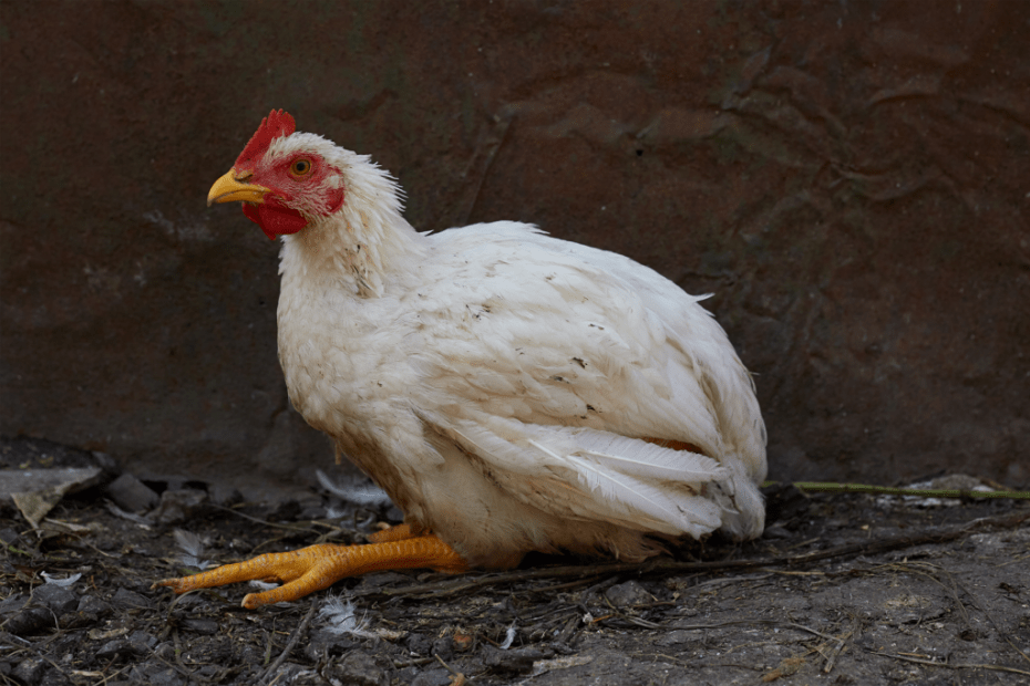
Coccidiosis in chickens can be a potentially life-threatening illness in your backyard flock. Chicks are particularly susceptible to coccidia, but chickens of any age can become ill. Coccidiosis disease affects chicks at an early age between 3 – 4weeks old or more depending on the type involved.
1. What is Coccidiosis?
Coccidiosis is caused by a tiny protozoan parasite called coccidia. Coccidia can only be seen with a microscope, but are bigger than bacteria.
They can be found naturally in the environment and there are hundreds of different types of coccidia and each one infects a different species of animal. Chickens have 7 different coccidia species that can infect them.
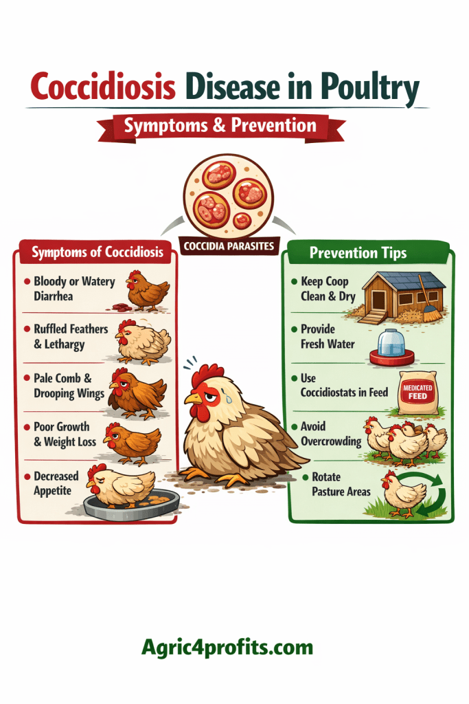
2. Symptoms of Coccidiosis Disease
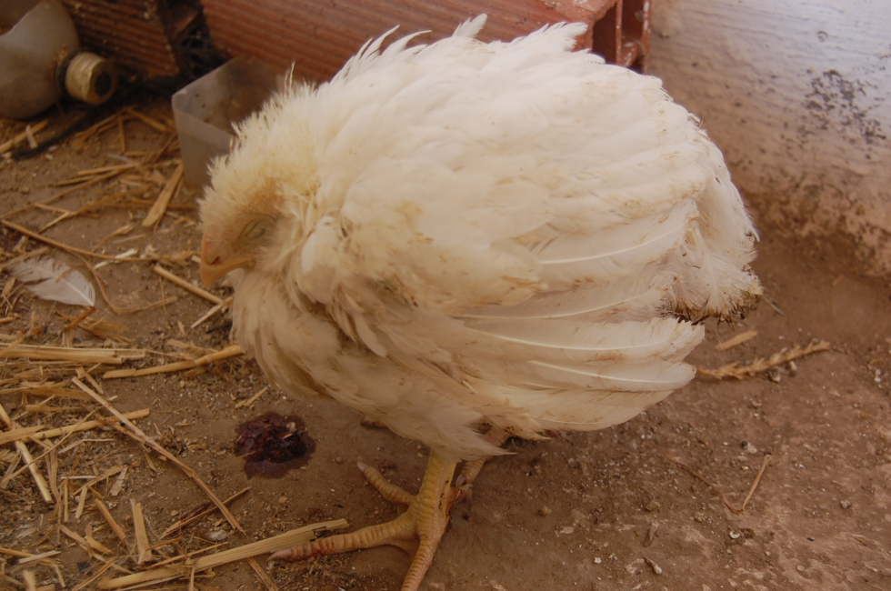
Chicks affected by coccidiosis disease will always close their eyes with feathers flying over and will not be able to feed or drink well as usual. There may also be bloody droppings, pale comb and ceasation of egg production in layers.
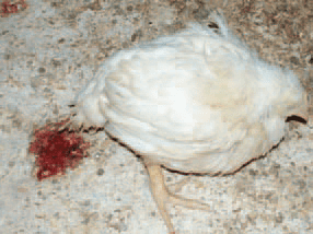
Coccidia causes illness in chickens by attacking the intestinal lining, causing diarrhea (sometimes bloody, but not always) which then leads to dehydration and malnutrition, and eventually death if not caught and treated immediately.
In some cases, the damage to the intestinal lining can be permanent and the bird may not grow or maintain body weight. Since a chick must eat a coccidia cyst to be infected, it takes a minimum of 2 to 3 weeks for a chick to begin showing symptoms. General lack of vigor or inactivity is usually the first sign, followed by loose, watery stools
3. Clinical Signs of Coccidiosis Disease
Coccidia which are deep tissue invaders such as E. maxima, E. necatrix and E. tenella cause severe necrosis, haemorrhage of the intestinal mucosa, and bloody diarrhoea and may result in death.
Signs include watery and/or bloody droppings, mortality (0-50%), and morbidity (0-100%). Culls appear as pale birds with anaemia, depression, poor weight gain and feed conversion, and a drop in egg production.
4. Prevention of Coccidiosis Disease in Chickens
Poultry farmers should ensure that litter are dried at all times. There should not be spillage of water on the floor. Good brooder hygiene is the best way to prevent an outbreak of coccidia in your chicks. Keep the litter in your brooder dry at all times. Replace the litter if it becomes saturated.
Since newly hatched chicks must be kept at a warmer temperature, the brooder is potentially the perfect environment to allow coccidia to explode in population and quickly infect your chicks. Many chicken keepers prefer to use anti-coccidiosis medications to help prevent a coccidiosis outbreak in their chicks.
One of the best ways to prevent a coccidiosis outbreak is by practicing responsible sanitation and litter management. Coccidia thrive in damp, warm conditions, so wet litter around the waterer is a virtual parasite paradise.
Believe it or not, when the conditions are just right, coccidia can survive for up to four years outside a bird’s body. And these hardy little organisms can be transmitted via boots, equipment, insects and rodents. So you’re going to need a multi-tiered approach to minimize the threat. Here are some suggestions:
- Keep the premises as dry as possible. Coccidia love moisture.
- Never introduce new adult birds into your flock. Birds that appear healthy can be carriers of a number of deadly diseases. Quarantine them first.
- Raise chicks in isolation. Mature birds can pass along diseases and parasites to vulnerable young birds.
- Thoroughly clean and disinfect the brooder between broods. This includes any equipment the chicks will come in contact with. Once the premises are dry, place four to six inches of dry, fresh litter material (wood shavings or a commercial absorbent litter material) on the floor.
- Provide clean water at all times. A typical problem is that brooder bedding or dust (containing feces) gets scratched into the water source. If possible, elevate the waterer slightly. Clean waterers relentlessly. If you wouldn’t be willing to drink the water yourself, it’s not clean enough. And never let the waterer run dry—it will force the birds to search for water in puddles, which are almost certainly contaminated.
- Provide clean bedding. Coccidia are spread through the feces of infected birds. If feces are in the bedding, they’re on the birds’ feathers. And if feces are on the feathers, the birds will ingest them while preening (using their beaks to clean themselves). Replace wet bedding around waterers and add bedding to any problem spots.
- Let sunlight do some of the work. Coccidia hate sunlight. It’s a natural disinfectant. Incorporate as much natural sunlight into your brooder as possible.
- Ask your veterinarian about vaccinating. A commercial coccidiosis vaccine is available, but it’s not beneficial for every flock. Consult your veterinarian before using the vaccine.
Remember, exposure to coccidia isn’t the threat—frankly, it’s unavoidable. Even wild birds carry coccidia. Instead, the serious threat comes from prolonged over-exposure to coccidia in a stressful, unsanitary environment that can overwhelm a bird’s immune system.
5. Treatment of Coccidiosis Disease in Chickens

If your chicks show signs of coccidiosis, you want to treat them immediately. To help reduce the coccidia population and limit the exposure to your chicks, do a complete change of bedding.
Also empty and disinfect all drinkers and feeders with a 10% bleach in water solution before beginning the treatment with an anti-coccidiosis medication. Treat the entire group of chicks by adding the medication to their drinking water according to label directions.
Read Also: Newcastle Disease: Symptoms and Prevention
6. List of Symptoms/Signs of Coccidiosis Disease
| Sign | Life Stages | Type |
|---|---|---|
| Cardiovascular Signs / Tachycardia, rapid pulse, high heart rate | Sign | |
| Cardiovascular Signs / Tachycardia, rapid pulse, high heart rate | Sign | |
| Digestive Signs / Abdominal distention | Sign | |
| Digestive Signs / Abnormal colour of stool in birds, white, green, yellow faeces | Cattle & Buffaloes:Calf,Poultry:Young poultry,Poultry:Mature female,Poultry:Cockerel,Poultry:Mature male,Other:All Stages,Pigs:Weaner,Pigs:Growing-finishing pig,Sheep & Goats:Lamb | Diagnosis |
| Digestive Signs / Anorexia, loss or decreased appetite, not nursing, off feed | Poultry:Young poultry,Poultry:Mature female,Poultry:Cockerel,Poultry:Mature male | Sign |
| Digestive Signs / Bloody stools, faeces, haematochezia | Poultry:Young poultry,Poultry:Mature female,Poultry:Cockerel,Poultry:Mature male | Diagnosis |
| Digestive Signs / Dark colour stools, faeces | Poultry:Young poultry,Poultry:Mature female,Poultry:Cockerel,Poultry:Mature male | Diagnosis |
| Digestive Signs / Diarrhoea | Cattle & Buffaloes:Calf,Poultry:Young poultry,Poultry:Mature female,Poultry:Cockerel,Poultry:Mature male,Other:All Stages,Pigs:Weaner,Pigs:Growing-finishing pig,Sheep & Goats:Lamb | Diagnosis |
| Digestive Signs / Excessive salivation, frothing at the mouth, ptyalism | Sign | |
| Digestive Signs / Hepatosplenomegaly, splenomegaly, hepatomegaly | Other:Adult Female,Other:Adult Male | Diagnosis |
| Digestive Signs / Melena or occult blood in faeces, stools | Poultry:Young poultry,Poultry:Mature female,Poultry:Cockerel,Poultry:Mature male,Other:Juvenile | Diagnosis |
| Digestive Signs / Mucous, mucoid stools, faeces | Cattle & Buffaloes:Calf,Poultry:Young poultry,Poultry:Mature female,Poultry:Cockerel,Poultry:Mature male,Pigs:Weaner,Pigs:Growing-finishing pig,Sheep & Goats:Lamb | Diagnosis |
| Digestive Signs / Parasites passed per rectum, in stools, faeces | Cattle & Buffaloes:All Stages,Poultry:All Stages,Other:All Stages,Pigs:All Stages,Sheep & Goats:All Stages | Diagnosis |
| Digestive Signs / Prolapsed rectum, rectal eversion | Sign | |
| Digestive Signs / Prolapsed rectum, rectal eversion | Sign | |
| Digestive Signs / Steatorrhea, fatty stools, faeces | Sign | |
| Digestive Signs / Sunken, empty crop in birds | Poultry:Young poultry,Poultry:Mature female,Poultry:Cockerel,Poultry:Mature male | Sign |
| Digestive Signs / Unusual or foul odor, stools, faeces | Cattle & Buffaloes:Calf,Poultry:Young poultry,Poultry:Mature female,Poultry:Cockerel,Poultry:Mature male,Other:All Stages,Pigs:Weaner,Pigs:Growing-finishing pig,Sheep & Goats:Lamb | Diagnosis |
| Digestive Signs / Vomiting or regurgitation, emesis | Sign | |
| General Signs / Ataxia, incoordination, staggering, falling | Sign | |
| General Signs / Ataxia, incoordination, staggering, falling | Sign | |
| General Signs / Dehydration | Cattle & Buffaloes:Calf,Poultry:Young poultry,Poultry:Mature female,Poultry:Cockerel,Poultry:Mature male,Other:All Stages,Pigs:Weaner,Pigs:Growing-finishing pig,Sheep & Goats:Lamb | Sign |
| General Signs / Dysmetria, hypermetria, hypometria | Sign | |
| General Signs / Fever, pyrexia, hyperthermia | Sign | |
| General Signs / Fever, pyrexia, hyperthermia | Sign | |
| General Signs / Generalized weakness, paresis, paralysis | Sign | |
| General Signs / Generalized weakness, paresis, paralysis | Sign | |
| General Signs / Inability to stand, downer, prostration | Sign | |
| General Signs / Inability to stand, downer, prostration | Sign | |
| General Signs / Inability to stand, downer, prostration | Sign | |
| General Signs / Increased mortality in flocks of birds | Sign | |
| General Signs / Lack of growth or weight gain, retarded, stunted growth | Sign | |
| General Signs / Lack of growth or weight gain, retarded, stunted growth | Sign | |
| General Signs / Lack of growth or weight gain, retarded, stunted growth | Sign | |
| General Signs / Opisthotonus | Sign | |
| General Signs / Pale comb and or wattles in birds | Poultry:Young poultry,Poultry:Mature female,Poultry:Cockerel,Poultry:Mature male | Sign |
| General Signs / Pale mucous membranes or skin, anemia | Poultry:Young poultry,Poultry:Mature female,Poultry:Cockerel,Poultry:Mature male | Sign |
| General Signs / Reluctant to move, refusal to move | Sign | |
| General Signs / Sudden death, found dead | Poultry:Young poultry,Poultry:Mature female,Poultry:Cockerel,Poultry:Mature male,Other:All Stages | Sign |
| General Signs / Tenesmus, straining, dyschezia | Sign | |
| General Signs / Tenesmus, straining, dyschezia | Sign | |
| General Signs / Trembling, shivering, fasciculations, chilling | Sign | |
| General Signs / Underweight, poor condition, thin, emaciated, unthriftiness, ill thrift | Cattle & Buffaloes:Calf,Poultry:Young poultry,Poultry:Mature female,Poultry:Cockerel,Poultry:Mature male,Other:All Stages,Pigs:Weaner,Pigs:Growing-finishing pig,Sheep & Goats:Lamb | Sign |
| General Signs / Weakness, paresis, paralysis of the legs, limbs in birds | Sign | |
| General Signs / Weight loss | Cattle & Buffaloes:Calf,Poultry:Young poultry,Poultry:Mature female,Poultry:Cockerel,Poultry:Mature male,Other:All Stages,Pigs:Weaner,Pigs:Growing-finishing pig,Sheep & Goats:Lamb | Diagnosis |
| Musculoskeletal Signs / Forelimb spasms, myoclonus | Sign | |
| Musculoskeletal Signs / Hindlimb spasms, myoclonus | Sign | |
| Nervous Signs / Abnormal behavior, aggression, changing habits | Sign | |
| Nervous Signs / Constant or increased vocalization | Sign | |
| Nervous Signs / Dullness, depression, lethargy, depressed, lethargic, listless | Poultry:Young poultry,Poultry:Mature female,Poultry:Cockerel,Poultry:Mature male,Other:Adult Female,Other:Adult Male | Sign |
| Nervous Signs / Excessive or decreased sleeping | Poultry:Young poultry,Poultry:Mature female,Poultry:Cockerel,Poultry:Mature male,Other:Adult Female,Other:Adult Male | Sign |
| Nervous Signs / Excitement, delirium, mania | Sign | |
| Nervous Signs / Head tilt | Sign | |
| Nervous Signs / Hyperesthesia, irritable, hyperactive | Sign | |
| Nervous Signs / Seizures or syncope, convulsions, fits, collapse | Sign | |
| Nervous Signs / Tetany | Sign | |
| Nervous Signs / Tremor | Sign | |
| Ophthalmology Signs / Blindness | Sign | |
| Ophthalmology Signs / Nystagmus | Sign | |
| Ophthalmology Signs / Strabismus | Sign | |
| Pain / Discomfort Signs / Pain, kidney, ureters, on palpation | Other:All Stages | Sign |
| Reproductive Signs / Decreased hatchability of eggs | Sign | |
| Reproductive Signs / Decreased, dropping, egg production | Sign | |
| Reproductive Signs / Male infertility | Sign | |
| Respiratory Signs / Dyspnea, difficult, open mouth breathing, grunt, gasping | Sign | |
| Respiratory Signs / Dyspnea, difficult, open mouth breathing, grunt, gasping | Sign | |
| Respiratory Signs / Increased respiratory rate, polypnea, tachypnea, hyperpnea | Sign | |
| Respiratory Signs / Increased respiratory rate, polypnea, tachypnea, hyperpnea | Sign | |
| Skin / Integumentary Signs / Rough hair coat, dull, standing on end | Sign | |
| Skin / Integumentary Signs / Rough hair coat, dull, standing on end | Sign | |
| Skin / Integumentary Signs / Ruffled, ruffling of the feathers | Sign |
Summary on Coccidiosis Disease: Symptoms and Prevention
| Aspect | Details |
|---|---|
| Disease Name | Coccidiosis |
| Definition | A common, highly prevalent protozoal parasitic disease of poultry caused by microscopic parasites that invade and destroy intestinal cells, causing enteritis, diarrhea, and significant production losses |
| Causative Agent | Protozoan parasites of the genus Eimeria (phylum Apicomplexa, family Eimeriidae); not bacteria or viruses |
| Eimeria Species in Chickens | Nine species described: E. acervulina, E. brunetti, E. hagani, E. maxima, E. mitis, E. mivati, E. necatrix, E. praecox, and E. tenella; the “big three” causing greatest economic losses are E. acervulina, E. maxima, and E. tenella |
| Eimeria Species in Turkeys | Several species including E. adenoides, E. gallopavonis, and E. meleagrimitis (the “big three” in turkeys); also affects ducks, geese, quail, and pheasants |
| Host Specificity | Strictly host-specific; Eimeria species infecting chickens do not infect turkeys, other livestock, pets, or humans; cross-infection between species does not occur |
| Intestinal Site Specificity | Each Eimeria species preferentially infects a specific segment of the intestinal tract; some infect the duodenum, others the mid-intestine, cecum, or entire intestinal tract |
| Transmission | Fecal-oral route; birds ingest sporulated oocysts (infective eggs) from contaminated litter, feed, water, soil, and feces; oocysts can be transported on clothing, footwear, equipment, and by insects |
| Life Cycle | Ingestion of sporulated oocysts → release of sporozoites → invasion of intestinal epithelial cells → multiple rounds of asexual replication (schizogony) → sexual cycle → new oocysts shed in feces; complete cycle takes approximately 4-7 days |
| Oocyst Sporulation | Freshly shed oocysts are non-infective; they sporulate and become infective within 1-2 days under warm, moist, and oxygenated conditions |
| Oocyst Persistence | Highly resistant to most common disinfectants; survive months to years in soil and litter; complete elimination from a production site is virtually impossible |
| Conditions Favoring Outbreaks | Wet or damp litter, overcrowding, high stocking density, poor ventilation, stress (cold, transport, diet change), immunosuppression (IBD, Marek’s disease), and introduction of new Eimeria species to a naive flock |
| Most Susceptible Age | Young birds aged 3-6 weeks; all ages susceptible but older birds with prior exposure develop partial immunity; battery-caged birds with no fecal contact rarely develop the disease |
| Disease Severity Factors | Eimeria species involved, infectious dose (oocyst load), host immune status, flock density, environmental conditions, concurrent infections (e.g., Clostridium perfringens causing necrotic enteritis) |
| Key Clinical Signs | Watery, mucoid, or bloody diarrhea; ruffled feathers; depression and lethargy; huddling; pale combs and wattles (anemia); dehydration; weight loss; reduced feed and water intake; wet vent area stained with blood or feces |
| Subclinical Form | Reduced growth rate, poor feed conversion ratio (FCR), decreased weight uniformity, and slightly reduced egg production with no obvious clinical signs; often more economically damaging than clinical outbreaks |
| Bloody Diarrhea | Characteristic of cecal coccidiosis (E. tenella) and small intestinal coccidiosis (E. necatrix); dark red to bright red blood passed in feces is a classic sign of severe infection |
| Morbidity Rate | Can reach 100% in susceptible naive flocks |
| Mortality Rate | Varies by species and dose; low in subclinical infections; up to 50-80% or higher in severe E. tenella or E. necatrix infections in unprotected flocks |
| Post-mortem Lesions | Intestinal wall thickening, hemorrhage, and necrosis; petechiae and blood clots in the cecum (E. tenella); ballooning and orange-red mucus in small intestine (E. maxima); white plaques or transverse striations in duodenum (E. acervulina); caseous cores in intestinal lumen |
| Pathological Mechanism | Parasite replication ruptures intestinal epithelial cells → destruction of intestinal mucosa → malabsorption of nutrients, protein loss, reduced vitamin A and K absorption, increased permeability, and secondary bacterial invasion |
| Secondary Complications | Necrotic enteritis (Clostridium perfringens overgrowth in damaged intestine), bacterial septicemia, E. coli infections; coccidiosis is the leading predisposing factor for necrotic enteritis |
| Diagnosis | Clinical signs + lesion scoring at necropsy (Johnson and Reid scoring system); confirmed by fecal flotation (oocyst detection), intestinal mucosal scraping microscopy, histopathology, or quantitative PCR (qPCR); oocyst count (OPG) helps determine severity |
| Differential Diagnosis | Necrotic enteritis, ulcerative enteritis, histomonosis (blackhead disease), infectious bursal disease, hemorrhagic enteritis, and nutritional deficiencies |
| Treatment | Anticoccidial drugs administered in drinking water (preferred during outbreaks for speed): amprolium, sulfadimethoxine, sulfaquinoxaline, toltrazuril (Baycox), diclazuril; supportive care with vitamins A and K aids intestinal recovery; sulfonamides NOT suitable for laying hens |
| Treatment Limitation | Drugs cannot completely halt an established outbreak; most intestinal damage occurs before clinical signs appear; prophylactic use is always preferable to treatment |
| Anticoccidial Drug Classes | Synthetic coccidiostats (decoquinate, diclazuril, halofuginone, nicarbazin, robenidine) and polyether ionophores (lasalocid, monensin, maduramicin, narasin, salinomycin, semduramicin); ionophores toxic to turkeys |
| Drug Resistance | Prolonged continuous use of the same anticoccidial promotes resistant Eimeria strains; rotation of anticoccidial programs is recommended to delay resistance development |
| Vaccination | Live attenuated (precocious) or non-attenuated oocyst vaccines given at hatch or day 1 (in ovo, spray, or drinking water); induces gradual natural immunity; replaces drug-resistant field strains with drug-sensitive vaccine strains; Paracox (7 species) widely used; vaccinated birds must NOT receive anticoccidial feed additives |
| Immunity | Species-specific; immunity to one Eimeria species does not protect against others; natural immunity develops after gradual exposure and requires 2-3 cycles of infection; immunity is maintained by low-level continuous field challenge |
| Litter Management | Maintaining dry, well-aerated litter reduces oocyst sporulation and build-up; avoid wet patches, overdrinking, and leaking drinkers; regular litter turning or replacement between flocks reduces oocyst load |
| Biosecurity | Footbaths, clothing changes, equipment disinfection (ammonia-based or formaldehyde disinfectants have some efficacy), all-in/all-out management, and preventing wild bird or insect access reduce introduction of new Eimeria strains |
| Economic Impact | Ranked the number one disease in the global broiler industry (2023 American Veterinarians in Broiler Production survey); annual global losses estimated at over $3 billion from reduced growth, mortality, medication costs, and secondary disease predisposition |
Frequently Asked Questions About Coccidiosis Disease: Symptoms and Prevention
1. What exactly causes coccidiosis in poultry and how is it different from bacterial or viral diseases?
Coccidiosis is caused by protozoan parasites of the genus Eimeria, making it a parasitic disease rather than a bacterial or viral one. Unlike bacteria or viruses, Eimeria are single-celled organisms with a complex multi-stage life cycle that takes place partly inside the host’s intestinal cells and partly in the environment. This distinction matters because the treatments, preventive drugs, and vaccines used for coccidiosis are completely different from antibiotics or antiviral agents. Standard disinfectants used against bacteria and viruses are largely ineffective against Eimeria oocysts.
2. How do chickens become infected with coccidiosis?
Infection occurs when a bird ingests sporulated oocysts (the infective stage of the parasite) from contaminated litter, soil, water, or feed. Oocysts are shed in the feces of infected or carrier birds and become infective (sporulate) within 1-2 days under warm, moist conditions. Because oocysts are present in virtually every poultry environment, exposure is essentially inevitable for birds raised on litter. The key factor determining whether exposure leads to disease is the dose of oocysts ingested relative to the bird’s level of immunity.
3. Why do some flocks develop coccidiosis even when they are on medicated feed?
Several factors can cause coccidiosis breakthrough even on medicated feeds. The most common reason is the development of drug-resistant Eimeria strains through prolonged use of the same anticoccidial compound without rotation. Other causes include underdosing (birds not consuming sufficient medicated feed due to heat stress or feed refusal), using medicated feed containing ingredients other than the intended anticoccidial (such as probiotics marketed as “medicated”), wet litter conditions creating an overwhelming oocyst challenge, and concurrent immunosuppressive diseases such as Infectious Bursal Disease or Marek’s Disease reducing the bird’s immune response.
4. What is the difference between subclinical and clinical coccidiosis, and which causes more economic damage?
Clinical coccidiosis produces obvious signs such as bloody diarrhea, high mortality, and birds visibly sick. Subclinical coccidiosis produces no visible illness but causes reduced growth rate, poor feed conversion, weight unevenness, and slightly reduced egg production. Subclinical coccidiosis is widely regarded as more economically damaging overall because it goes undetected for longer, affects entire flocks continuously, and its cumulative production losses (feed wasted, reduced weight gain, increased days to market) far exceed those of dramatic but short-lived clinical outbreaks that are quickly recognized and treated.
5. Can coccidiosis infect humans or other animals on the farm?
No. Eimeria species are strictly host-specific, meaning the species that infect chickens will not infect turkeys, ducks, other livestock, pets, or humans. This is a fundamental biological characteristic of the parasite. However, humans, clothing, boots, and equipment can physically carry oocysts between flocks and houses, making farm workers an important vehicle for spreading the disease between pens and farms even though they cannot themselves become infected.
6. How does coccidiosis vaccination work and is it better than using anticoccidial drugs?
Coccidiosis vaccines contain live Eimeria oocysts, either attenuated (weakened by precocious selection) or non-attenuated (full-virulence). When administered at hatch or day 1, the vaccine oocysts undergo a limited cycle of replication in the gut, stimulating a natural immune response. Repeated field exposure through normal litter contact then boosts and maintains this immunity. Vaccination is particularly valuable in long-lived flocks (breeders, layers), antibiotic-free and organic production systems, and situations where drug resistance has become a significant problem. The main advantage of vaccination over drugs is that it establishes sustainable, naturally renewed immunity. The main disadvantage is that immunity takes 3-4 weeks to develop fully, leaving young chicks temporarily vulnerable.
7. Why are ionophore anticoccidial drugs toxic to turkeys but safe for chickens?
Turkeys are inherently sensitive to polyether ionophore antibiotics (monensin, lasalocid, salinomycin, narasin, maduramicin) at doses that are safe and effective in chickens. The exact mechanism of this species difference involves how turkeys metabolize these compounds and their greater cardiac sensitivity to ionophore-induced ion transport disruption. Accidental feeding of ionophore-containing chicken feed to turkeys can cause leg weakness, paralysis, heart failure, and death. This is a critical practical concern in mixed-species poultry operations, and all feed bags must be carefully checked for ionophore content before being given to turkeys.
8. How do I know if my birds have coccidiosis or necrotic enteritis, since the signs can look similar?
Both diseases cause intestinal damage, diarrhea, depression, and mortality, and they frequently occur together since coccidiosis is the primary predisposing factor for necrotic enteritis. Key differences: coccidiosis typically shows bloody or mucoid diarrhea with hemorrhagic intestinal lesions and visible oocysts on microscopy; necrotic enteritis (caused by Clostridium perfringens) produces a characteristic “Turkish towel” appearance of the small intestinal mucosa with a foul-smelling necrotic membrane, and bacteria are seen on smear. Mixed infections are common and require both anticoccidial treatment and appropriate antibiotics. A veterinarian and laboratory confirmation are advisable for accurate differentiation.
9. How long do Eimeria oocysts survive in the environment and how can I reduce their numbers between flocks?
Eimeria oocysts are exceptionally resistant and can survive for months to over a year in moist soil and litter, even at low temperatures. They are resistant to most common disinfectants. Complete eradication from a production site is virtually impossible. However, their numbers can be significantly reduced by complete removal and composting of used litter between flocks, thorough cleaning and drying of housing (desiccation kills oocysts), application of ammonia-releasing compounds (quicklime) or formaldehyde-based disinfectants, and maintaining dry conditions throughout the flock cycle. A downtime period of at least 2 weeks between flocks, combined with litter cleanout, substantially reduces the oocyst challenge for the incoming flock.
10. What are the most important steps to prevent coccidiosis on a poultry farm?
A comprehensive coccidiosis prevention program rests on four pillars. First, use either a properly chosen and rotated anticoccidial drug program in feed (remembering that birds on vaccination must not receive anticoccidials) or a licensed coccidiosis vaccine administered correctly at hatch. Second, maintain dry litter through good ventilation, proper drinker management, and prompt replacement of wet patches. Third, apply all-in/all-out management with thorough cleanout and downtime between flocks. Fourth, avoid or manage risk factors including overcrowding, immunosuppressive diseases, stress, and sudden diet changes that lower the bird’s natural resistance and allow overwhelming oocyst challenge to cause disease.
Read Also: Low-Maintenance Plants for Beginners