
The life cycle of plants alternates between a multicellular haploid gametophyte and a multicellular diploid sporophyte, both differing morphologically and functionally.
According to Gifford and Foster (1989), in the life cycle of a plant:
i. Diploid sporophyte produces haploid spores through meiosis.
ii. Spores develop into gametophytes.
iii. Gametophytes produce haploid gametes.
iv. The fusion of the female gamete (egg) and the male gamete results in the formation of a zygote.
v. The zygote marks the beginning of the diploid sporophyte phase.
In Angiosperms, the diploid sporophyte produces two types of spores: microspores and megaspores. The microspore develops into the male gametophyte, and the megaspore produces the female gametophyte, which will be explored in this article.
The gametophyte in Angiosperms develops within sporophytic tissue, specifically in the sexual organs of the flower. The male gametophyte (pollen grain, composed of two sperm cells within a vegetative cell) develops in the anther, the fertile part of the stamen (McCormick, 1993, 2004). The female gametophyte (embryo sac) develops within the ovule, found in the ovary.
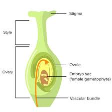
The gynoecium or pistil represents the female reproductive organ in a flower, while the carpel is a unit of the pistil. Each carpel consists of a basal swollen ovary containing one or more ovules, a receptive stigma, and often a stalk-like style between them.
Ovules, enclosed by the ovary wall, are attached to the carpellary tissue known as the placenta. The arrangement of ovules within the ovary is termed placentation.
The ovule (or megasporangium) is the site where megaspores and the female gametophyte form. After fertilization, the female gametophyte produces the embryo and endosperm, while the entire megasporangium with its enclosed structure develops into the seed, ensuring the continuity of the next generation.
Read Also: Coccidiosis Disease in Poultry: Symptoms and Prevention
Structure of the Ovule in Angiosperms
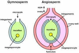
The megasporangium or ovule consists of the nucellus and its protective coverings, known as integuments. It is attached to the placenta within the ovary by a stalk called the funiculus. The point of attachment of the ovule to the funiculus is termed the hilum.
A mature ovule, ready for fertilization, contains the nucellus, which is almost entirely covered by one or two sheaths (integuments), leaving a small opening at the apical end known as the micropyle.
The basal region of the ovule, where it attaches to the placenta by the funiculus, is called the chalaza, and this side is referred to as the chalazal end. The opposite end is the micropylar end, which serves as the passage for the entry of the pollen tube into the ovule. Inside the nucellus lies the female gametophyte, also known as the embryo sac.
The nucellar tissue, which is parenchymatous, represents the wall of the megasporangium. The nucellus is mostly consumed by the developing embryo sac or endosperm. Each ovule typically contains a single nucellus, although two nuclei may sometimes occur as an abnormality, as observed in Aegle marmelos (Fig. 9.8).
The ovule with one integument is called unitegmic, with two integuments is bitegmic, and without integument is referred to as ategmic.
Parts of the Ovule
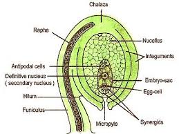
1. Funiculus or Funicle: Stalk attaching the ovule to the placenta
2. Nucellus: The body of the ovule.
3. Integument: Protective covering of the nucellus.
4. Micropyle: Small opening formed by the integuments at the tip of the nucellus.
5. Chalaza: The basal part of the ovule
6. Hilum: Region where the ovule fuses with te funiculus.
7. Embryo sac: Female gametophyte located within the nucellus, developed from the megaspore.
Types of Ovules in Plants
Based on the position of the micropyle concerning the funiculus, mature ovules are classified into six main types:
1. Orthotropous: The ovule is straight, with the micropyle positioned in line with the funiculus.
2. Anatropous: The ovule is inverted, making the micropyle point towards the funiculus.
3. Campylotropous: The ovule is curved, causing the micropyle to be bent towards the funiculus.
4. Amphitropous: The ovule is partially inverted, with both the body and funiculus showing a curved shape, making the micropyle and funiculus nearly parallel.
5. Hemianatropous: The ovule is half-inverted, where the micropyle takes a slightly inclined position towards the funiculus.
6. Circinotropous: The ovule undergoes a full rotation during development, making the micropyle point towards the funiculus in a circular arrangement.
Ovules can also be categorized based on the development of the nucellus and the position of the sporogenous cell:
1. Tenuinucellate type: The archesporial cell directly functions as the megaspore mother cell (MMC), and the nucellar tissue remains single-layered.
2. Crassinucellate type: The archesporial cell divides into a parietal cell and a sporogenous cell, embedding the sporogenous cell in a massive nucellus.
Read Also: Newcastle Disease in Poultry: Symptoms and Prevention
Development of the Ovule in Plants
The ovule first arises as a primordium on the placenta within the ovary cavity. Due to the meristematic activity of the ovular primordia cells, the protuberance grows into a mass of tissue called the nucellus. Two integuments develop at the base of the nucellus, and the ovule assumes its final shape during the megaspore tetrad stage
The development of the female gametophyte occurs in two phases:
1. Megasporogenesis: This phase involves the formation of megaspores from a megaspore mother cell through the process of meiosis. Out of the four megaspores produced, usually, only one remains functional, while the others degenerate.
2. Megagametogenesis: In this phase, the functional megaspore develops into the embryo sac (female gametophyte) through mitotic divisions, resulting in the formation of the egg cell and other supporting cells within the ovule.
The most common developmental pattern, referred to as the Polygonum type, was first described in Polygonum divaricatum (Strasburger, 1879; Maheshwari, 1950).
Megasporogenesis in Ovules
Megasporogenesis refers to the development of the megaspore within the ovule. A hypodermal cell at the micropylar end differentiates and functions as the archesporium.
The megaspore mother cell (MMC) undergoes meiosis, producing four haploid megaspores in a linear tetrad. One megaspore remains functional, while the other three degenerate.
Pollination in Angiosperms
The next biological phase following male and female gametophyte development is pollination. Pollination is the transfer of pollen from the anther to the receptive stigma, either within the same flower or between different flowers.
Two main types of pollination occur:
1. Self-pollination (autogamy): Transfer of pollen to the stigma of the same flower or another flower on the same plant, as in peas, wheat, and rice.
2. Cross-pollination (allogamy): Transfer of pollen to the stigma of a different flower, as in hemp and willow.
Pollination ensures fertilization, leading to the formation of seeds and fruits, thereby continuing the plant’s life cycle.
Agents of Pollination in Agriculture
Pollination occurs through various agents, classified into:
1. Abiotic agents: Wind (anemophily) and water (hydrophily).
2. Biotic agents: Insects (entomophily), birds (ornithophily), and bats (cheiropterophily).
In this article, the structure, types, and development of ovules were discussed. The next phase after gametophyte development is pollination, which is crucial for fertilization and the production of seeds. Pollination is facilitated by abiotic (wind and water) and biotic (insects, birds, bats) agents, ensuring the continuation of plant life.
Do you have any questions, suggestions, or contributions? If so, please feel free to use the comment box below to share your thoughts. We also encourage you to kindly share this information with others who might benefit from it. Since we can’t reach everyone at once, we truly appreciate your help in spreading the word. Thank you so much for your support and for sharing!
Frequently Asked Questions
We will update this section soon.